6XCS | pdb_00006xcs

Erythromycin esterase mutant EreC H289N in its open conformation


Experimental Data Snapshot

  • Method: X-RAY DIFFRACTION
  • Resolution: 2.40 Å
  • R-Value Free: 
    0.290 (Depositor), 0.292 (DCC) 
  • R-Value Work: 
    0.240 (Depositor), 0.243 (DCC) 
  • R-Value Observed: 
    0.243 (Depositor) 

Starting Model: experimental
View more details

wwPDB Validation 3D Report Full Report

Validation slider image for 6XCS

This is version 1.4 of the entry. See complete history

Literature

Structural and functional insights into esterase-mediated macrolide resistance.

Zielinski, M.Park, J.Sleno, B.Berghuis, A.M.

(2021) Nat Commun 12: 1732-1732

  • DOI: https://doi.org/10.1038/s41467-021-22016-3
  • Primary Citation Related Structures: 
    6XCQ, 6XCS

  • PubMed Abstract: 

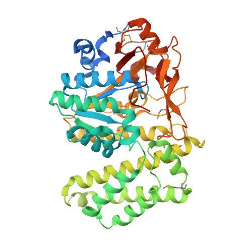
    Macrolides are a class of antibiotics widely used in both medicine and agriculture. Unsurprisingly, as a consequence of their exensive usage a plethora of resistance mechanisms have been encountered in pathogenic bacteria. One of these resistance mechanisms entails the enzymatic cleavage of the macrolides' macrolactone ring by erythromycin esterases (Eres). The most frequently identified Ere enzyme is EreA, which confers resistance to the majority of clinically used macrolides. Despite the role Eres play in macrolide resistance, research into this family enzymes has been sparse. Here, we report the first three-dimensional structures of an erythromycin esterase, EreC. EreC is an extremely close homologue of EreA, displaying more than 90% sequence identity. Two structures of this enzyme, in conjunction with in silico flexible docking studies and previously reported mutagenesis data allowed for the proposal of a detailed catalytic mechanism for the Ere family of enzymes, labeling them as metal-independent hydrolases. Also presented are substrate spectrum assays for different members of the Ere family. The results from these assays together with an examination of residue conservation for the macrolide binding site in Eres, suggests two distinct active site archetypes within the Ere enzyme family.


  • Organizational Affiliation
    • Department of Biochemistry, McGill University, Montréal, QC, Canada.

Macromolecule Content 

  • Total Structure Weight: 97.84 kDa 
  • Atom Count: 6,169 
  • Modeled Residue Count: 816 
  • Deposited Residue Count: 876 
  • Unique protein chains: 1

Macromolecules

Find similar proteins by:|  3D Structure
Entity ID: 1
MoleculeChains  Sequence LengthOrganismDetailsImage
EreC
A, B
438Klebsiella pneumoniaeMutation(s): 1 
Gene Names: ereC
UniProt
Find proteins for C7C425 (Klebsiella pneumoniae)
Explore C7C425 
Go to UniProtKB:  C7C425
Entity Groups
Sequence Clusters30% Identity50% Identity70% Identity90% Identity95% Identity100% Identity
UniProt GroupC7C425
Sequence Annotations
Expand
Reference Sequence

Small Molecules

Modified Residues  1 Unique
IDChains TypeFormula2D DiagramParent
MSE
Query on MSE
A, B
L-PEPTIDE LINKINGC5 H11 N O2 SeMET

Experimental Data & Validation

Experimental Data

  • Method: X-RAY DIFFRACTION
  • Resolution: 2.40 Å
  • R-Value Free:  0.290 (Depositor), 0.292 (DCC) 
  • R-Value Work:  0.240 (Depositor), 0.243 (DCC) 
  • R-Value Observed: 0.243 (Depositor) 
Space Group: C 1 2 1
Unit Cell:
Length ( Å )Angle ( ˚ )
a = 95.367α = 90
b = 74.214β = 96.498
c = 129.29γ = 90
Software Package:
Software NamePurpose
PHENIXrefinement
xia2data reduction
DIALSdata scaling
PHASERphasing

Structure Validation

View Full Validation Report


Entry History 

& Funding Information

Deposition Data


Funding OrganizationLocationGrant Number
Canadian Institutes of Health Research (CIHR)CanadaPJT-162365

Revision History  (Full details and data files)

  • Version 1.0: 2021-02-17
    Type: Initial release
  • Version 1.1: 2021-03-31
    Changes: Database references
  • Version 1.2: 2023-10-18
    Changes: Data collection, Database references, Refinement description
  • Version 1.3: 2023-11-15
    Changes: Data collection
  • Version 1.4: 2024-10-30
    Changes: Structure summary