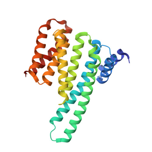
6XAG

Apo BRAF dimer bound to 14-3-3


Experimental Data Snapshot

  • Method: X-RAY DIFFRACTION
  • Resolution: 3.30 Å
  • R-Value Free: 0.300 
  • R-Value Work: 0.250 
  • R-Value Observed: 0.252 

Starting Model: experimental
View more details

wwPDB Validation   3D Report Full Report


This is version 1.3 of the entry. See complete history


Literature

Dimerization Induced by C-Terminal 14-3-3 Binding Is Sufficient for BRAF Kinase Activation.

Liau, N.P.D.Venkatanarayan, A.Quinn, J.G.Phung, W.Malek, S.Hymowitz, S.G.Sudhamsu, J.

(2020) Biochemistry 59: 3982-3992

  • DOI: https://doi.org/10.1021/acs.biochem.0c00517
  • Primary Citation of Related Structures:  
    6XAG

  • PubMed Abstract: 

    The Ras-RAF-MEK-ERK signaling axis, commonly mutated in human cancers, is highly regulated to prevent aberrant signaling in healthy cells. One of the pathway modulators, 14-3-3, a constitutive dimer, induces RAF dimerization and activation by binding to a phosphorylated motif C-terminal to the RAF kinase domain. Recent work has suggested that a C-terminal "DTS" region in BRAF is necessary for this 14-3-3-mediated activation. We show that the catalytic activity and ATP binding affinity of the BRAF:14-3-3 complex is insensitive to the presence or absence of the DTS, while the ATP sites of both BRAF molecules are identical and available for binding. We also present a crystal structure of the apo BRAF:14-3-3 complex showing that the DTS is not required to attain the catalytically active conformation of BRAF. Rather, BRAF dimerization induced by 14-3-3 is the key step in activation, allowing the active BRAF:14-3-3 tetramer to achieve catalytic activity comparable to the constitutively active oncogenic BRAF V600E mutant.


Macromolecules
Find similar proteins by:  (by identity cutoff)  |  3D Structure
Entity ID: 1
MoleculeChains Sequence LengthOrganismDetailsImage
14-3-3 protein zeta/delta
A, B
232Homo sapiensMutation(s): 0 
Gene Names: YWHAZ
UniProt & NIH Common Fund Data Resources
Find proteins for P63104 (Homo sapiens)
Explore P63104 
Go to UniProtKB:  P63104
PHAROS:  P63104
GTEx:  ENSG00000164924 
Entity Groups  
Sequence Clusters30% Identity50% Identity70% Identity90% Identity95% Identity100% Identity
UniProt GroupP63104
Sequence Annotations
Expand
  • Reference Sequence
Find similar proteins by:  (by identity cutoff)  |  3D Structure
Entity ID: 2
MoleculeChains Sequence LengthOrganismDetailsImage
Serine/threonine-protein kinase B-raf
C, D
290Homo sapiensMutation(s): 0 
Gene Names: BRAFBRAF1RAFB1
EC: 2.7.11.1
UniProt & NIH Common Fund Data Resources
Find proteins for P15056 (Homo sapiens)
Explore P15056 
Go to UniProtKB:  P15056
PHAROS:  P15056
GTEx:  ENSG00000157764 
Entity Groups  
Sequence Clusters30% Identity50% Identity70% Identity90% Identity95% Identity100% Identity
UniProt GroupP15056
Sequence Annotations
Expand
  • Reference Sequence
Small Molecules
Ligands 1 Unique
IDChains Name / Formula / InChI Key2D Diagram3D Interactions
EDO
Query on EDO

Download Ideal Coordinates CCD File 
E [auth D]1,2-ETHANEDIOL
C2 H6 O2
LYCAIKOWRPUZTN-UHFFFAOYSA-N
Modified Residues  1 Unique
IDChains TypeFormula2D DiagramParent
SEP
Query on SEP
C, D
L-PEPTIDE LINKINGC3 H8 N O6 PSER
Experimental Data & Validation

Experimental Data

  • Method: X-RAY DIFFRACTION
  • Resolution: 3.30 Å
  • R-Value Free: 0.300 
  • R-Value Work: 0.250 
  • R-Value Observed: 0.252 
  • Space Group: P 21 21 21
Unit Cell:
Length ( Å )Angle ( ˚ )
a = 61.504α = 90
b = 115.752β = 90
c = 153.092γ = 90
Software Package:
Software NamePurpose
PHENIXrefinement
PDB_EXTRACTdata extraction
XDSdata reduction
XDSdata scaling
PHENIXphasing

Structure Validation

View Full Validation Report


Entry History 

Deposition Data

Revision History  (Full details and data files)

  • Version 1.0: 2020-10-07
    Type: Initial release
  • Version 1.1: 2020-11-04
    Changes: Database references
  • Version 1.2: 2023-10-18
    Changes: Data collection, Database references, Refinement description
  • Version 1.3: 2024-10-23
    Changes: Structure summary