6X7H | pdb_00006x7h

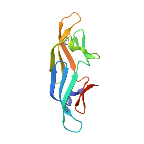
Cyanovirin-N Mutation I34Y with Dimannose bound


Experimental Data Snapshot

  • Method: X-RAY DIFFRACTION
  • Resolution: 1.25 Å
  • R-Value Free: 
    0.173 (Depositor), 0.173 (DCC) 
  • R-Value Work: 
    0.140 (Depositor), 0.141 (DCC) 
  • R-Value Observed: 
    0.141 (Depositor) 

Starting Model: experimental
View more details

wwPDB Validation 3D Report Full Report

Validation slider image for 6X7H

This is version 1.3 of the entry. See complete history

Literature

Design of novel cyanovirin-N variants by modulation of binding dynamics through distal mutations.

Kazan, I.C.Sharma, P.Rahman, M.I.Bobkov, A.Fromme, R.Ghirlanda, G.Ozkan, S.B.

(2022) Elife 11

  • DOI: https://doi.org/10.7554/eLife.67474
  • Primary Citation Related Structures: 
    6X7H

  • PubMed Abstract: 

    We develop integrated co-evolution and dynamic coupling (ICDC) approach to identify, mutate, and assess distal sites to modulate function. We validate the approach first by analyzing the existing mutational fitness data of TEM-1 β-lactamase and show that allosteric positions co-evolved and dynamically coupled with the active site significantly modulate function. We further apply ICDC approach to identify positions and their mutations that can modulate binding affinity in a lectin, cyanovirin-N (CV-N), that selectively binds to dimannose, and predict binding energies of its variants through Adaptive BP-Dock. Computational and experimental analyses reveal that binding enhancing mutants identified by ICDC impact the dynamics of the binding pocket, and show that rigidification of the binding residues compensates for the entropic cost of binding. This work suggests a mechanism by which distal mutations modulate function through dynamic allostery and provides a blueprint to identify candidates for mutagenesis in order to optimize protein function.


  • Organizational Affiliation
    • Center for Biological Physics and Department of Physics, Arizona State University, Tempe, United States.

Macromolecule Content 

  • Total Structure Weight: 24.68 kDa 
  • Atom Count: 1,885 
  • Modeled Residue Count: 200 
  • Deposited Residue Count: 218 
  • Unique protein chains: 1

Macromolecules

Find similar proteins by:|  3D Structure
Entity ID: 1
MoleculeChains  Sequence LengthOrganismDetailsImage
Cyanovirin-N
A, B
109Nostoc ellipsosporumMutation(s): 1 
UniProt
Find proteins for P81180 (Nostoc ellipsosporum)
Explore P81180 
Go to UniProtKB:  P81180
Entity Groups
Sequence Clusters30% Identity50% Identity70% Identity90% Identity95% Identity100% Identity
UniProt GroupP81180
Sequence Annotations
Expand
Reference Sequence

Oligosaccharides

Help  
Entity ID: 2
MoleculeChains Length2D Diagram GlycosylationD Interactions
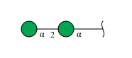
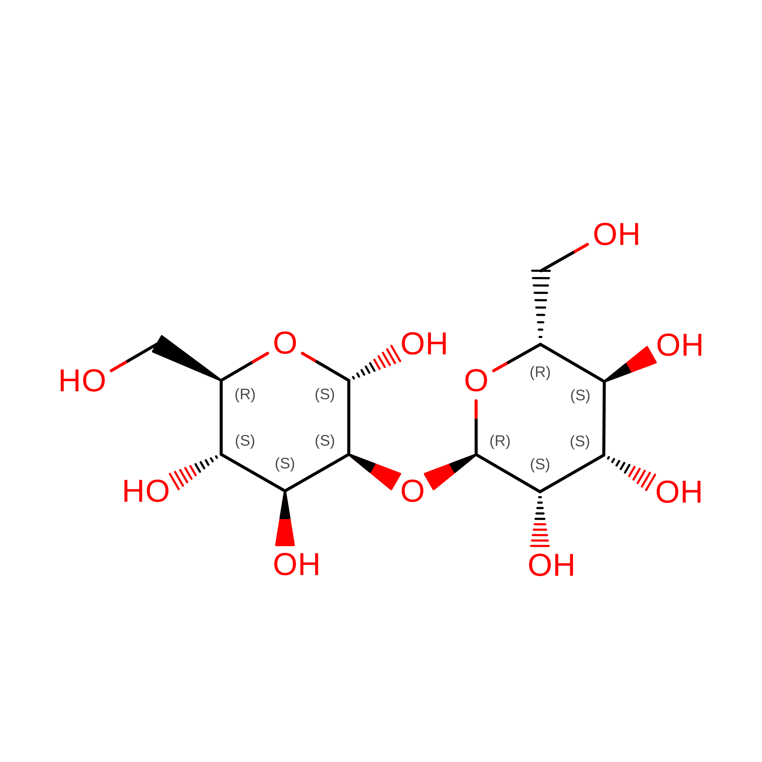
alpha-D-mannopyranose-(1-2)-alpha-D-mannopyranose
C, D
2N/A
Glycosylation Resources
GlyTouCan: G53402KW
GlyCosmos: G53402KW
GlyGen: G53402KW

Biologically Interesting Molecules (External Reference) 

1 Unique

Experimental Data & Validation

Experimental Data

  • Method: X-RAY DIFFRACTION
  • Resolution: 1.25 Å
  • R-Value Free:  0.173 (Depositor), 0.173 (DCC) 
  • R-Value Work:  0.140 (Depositor), 0.141 (DCC) 
  • R-Value Observed: 0.141 (Depositor) 
Space Group: P 1 21 1
Unit Cell:
Length ( Å )Angle ( ˚ )
a = 49.724α = 90
b = 38.011β = 100
c = 55.867γ = 90
Software Package:
Software NamePurpose
PHENIXrefinement
XDSdata reduction
Aimlessdata scaling
PHASERphasing

Structure Validation

View Full Validation Report


Entry History 

Deposition Data

Revision History  (Full details and data files)

  • Version 1.0: 2021-06-02
    Type: Initial release
  • Version 1.1: 2022-12-14
    Changes: Database references, Refinement description
  • Version 1.2: 2023-10-25
    Changes: Data collection, Refinement description
  • Version 1.3: 2024-11-06
    Changes: Structure summary