6X6Q | pdb_00006x6q

Peptide-bound structure of Marinomonas primoryensis peptide-binding domain


Experimental Data Snapshot

  • Method: X-RAY DIFFRACTION
  • Resolution: 2.17 Å
  • R-Value Free: 
    0.202 (Depositor), 0.200 (DCC) 
  • R-Value Work: 
    0.173 (Depositor), 0.170 (DCC) 
  • R-Value Observed: 
    0.175 (Depositor) 

Starting Model: experimental
View more details

wwPDB Validation   3D Report Full Report


This is version 1.2 of the entry. See complete history


Literature

Molecular basis for inhibition of adhesin-mediated bacterial-host interactions through a peptide-binding domain.

Guo, S.Zahiri, H.Stevens, C.Spaanderman, D.C.Milroy, L.G.Ottmann, C.Brunsveld, L.Voets, I.K.Davies, P.L.

(2021) Cell Rep 37: 110002-110002

  • DOI: https://doi.org/10.1016/j.celrep.2021.110002
  • Primary Citation of Related Structures:  
    6X5V, 6X5W, 6X6M, 6X6Q

  • PubMed Abstract: 

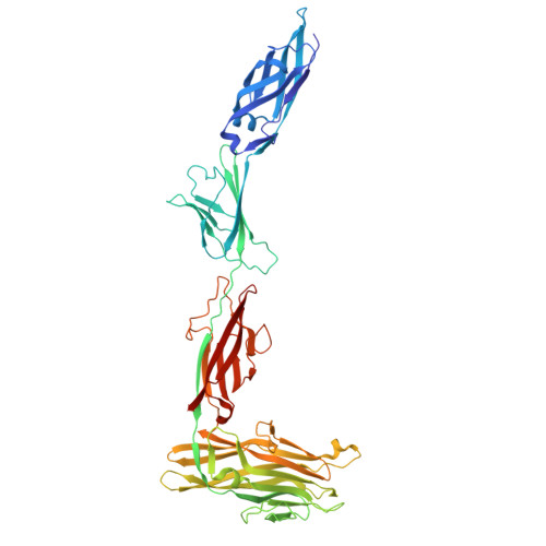
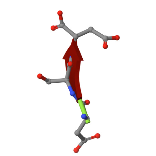
    Infections typically begin with pathogens adhering to host cells. For bacteria, this adhesion can occur through specific ligand-binding domains. We identify a 20-kDa peptide-binding domain (PBD) in a 1.5-MDa RTX adhesin of a Gram-negative marine bacterium that colonizes diatoms. The crystal structure of this Ca 2+ -dependent PBD suggests that it may bind the C termini of host cell-surface proteins. A systematic peptide library analysis reveals an optimal tripeptide sequence with 30-nM affinity for the PBD, and X-ray crystallography details its peptide-protein interactions. Binding of the PBD to the diatom partner of the bacteria can be inhibited or competed away by the peptide, providing a molecular basis for inhibiting bacterium-host interactions. We further show that this PBD is found in other bacteria, including human pathogens such as Vibrio cholerae and Aeromonas veronii. Here, we produce the PBD ortholog from A. veronii and demonstrate, using the same peptide inhibitor, how pathogens may be prevented from adhering to their hosts.


  • Organizational Affiliation
    • Department of Biomedical and Molecular Sciences, Queen's University, Kingston, ON K7L 3N6, Canada; Laboratory of Self-Organizing Soft Matter, Eindhoven University of Technology PO Box 513, 5600 MB Eindhoven, the Netherlands; Laboratory of Chemical Biology, Eindhoven University of Technology PO Box 513, 5600 MB Eindhoven, the Netherlands; Institute for Complex Molecular Systems, Eindhoven University of Technology PO Box 513, 5600 MB Eindhoven, the Netherlands.

Macromolecules
Find similar proteins by:  (by identity cutoff)  |  3D Structure
Entity ID: 1
MoleculeChains Sequence LengthOrganismDetailsImage
Antifreeze protein507Marinomonas primoryensisMutation(s): 0 
UniProt
Find proteins for A1YIY3 (Marinomonas primoryensis)
Explore A1YIY3 
Go to UniProtKB:  A1YIY3
Entity Groups  
Sequence Clusters30% Identity50% Identity70% Identity90% Identity95% Identity100% Identity
UniProt GroupA1YIY3
Sequence Annotations
Expand
  • Reference Sequence

Find similar proteins by:  Sequence   |   3D Structure  

Entity ID: 2
MoleculeChains Sequence LengthOrganismDetailsImage
ASP-SER-ASP3synthetic constructMutation(s): 0 
Sequence Annotations
Expand
  • Reference Sequence
Small Molecules
Ligands 2 Unique
IDChains Name / Formula / InChI Key2D Diagram3D Interactions
1PE
Query on 1PE

Download Ideal Coordinates CCD File 
S [auth A],
T [auth A]
PENTAETHYLENE GLYCOL
C10 H22 O6
JLFNLZLINWHATN-UHFFFAOYSA-N
CA
Query on CA

Download Ideal Coordinates CCD File 
C [auth A]
D [auth A]
E [auth A]
F [auth A]
G [auth A]
C [auth A],
D [auth A],
E [auth A],
F [auth A],
G [auth A],
H [auth A],
I [auth A],
J [auth A],
K [auth A],
L [auth A],
M [auth A],
N [auth A],
O [auth A],
P [auth A],
Q [auth A],
R [auth A]
CALCIUM ION
Ca
BHPQYMZQTOCNFJ-UHFFFAOYSA-N
Experimental Data & Validation

Experimental Data

  • Method: X-RAY DIFFRACTION
  • Resolution: 2.17 Å
  • R-Value Free:  0.202 (Depositor), 0.200 (DCC) 
  • R-Value Work:  0.173 (Depositor), 0.170 (DCC) 
  • R-Value Observed: 0.175 (Depositor) 
Space Group: P 1
Unit Cell:
Length ( Å )Angle ( ˚ )
a = 50.165α = 66.612
b = 60.717β = 72.536
c = 63.658γ = 68.582
Software Package:
Software NamePurpose
PHENIXrefinement
xia2data reduction
xia2data scaling
PHASERphasing

Structure Validation

View Full Validation Report


Entry History & Funding Information

Deposition Data


Funding OrganizationLocationGrant Number
Canadian Institutes of Health Research (CIHR)--

Revision History  (Full details and data files)

  • Version 1.0: 2021-09-01
    Type: Initial release
  • Version 1.1: 2024-04-03
    Changes: Data collection, Refinement description
  • Version 1.2: 2026-01-21
    Changes: Database references, Structure summary