6X5M | pdb_00006x5m

Crystal structure of a stabilized PAN ENE bimolecular triplex with a GC-clamped polyA tail, in complex with Fab-BL-3,6.


Experimental Data Snapshot

  • Method: X-RAY DIFFRACTION
  • Resolution: 2.50 Å
  • R-Value Free: 
    0.279 (Depositor), 0.283 (DCC) 
  • R-Value Work: 
    0.207 (Depositor), 0.210 (DCC) 
  • R-Value Observed: 
    0.209 (Depositor) 

Starting Model: experimental
View more details

wwPDB Validation 3D Report Full Report

Validation slider image for 6X5M

Ligand Structure Quality Assessment 


This is version 2.4 of the entry. See complete history

Literature

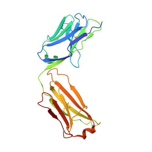
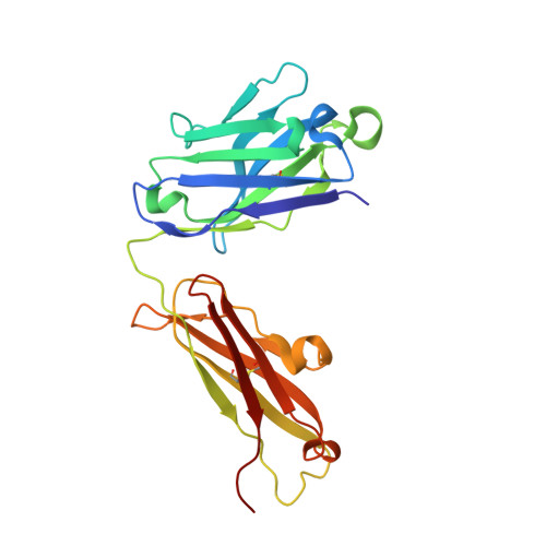
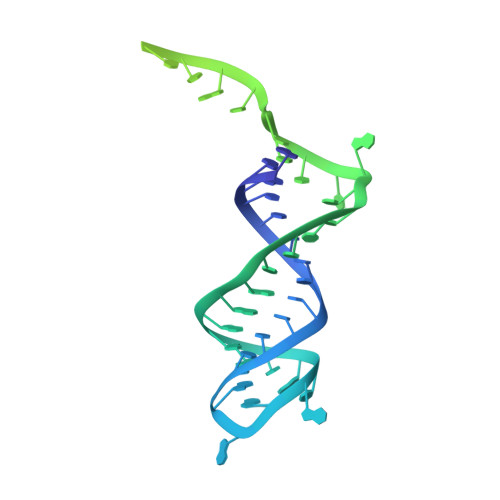
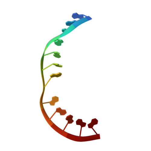
Dynamic bulge nucleotides in the KSHV PAN ENE triple helix provide a unique binding platform for small molecule ligands.

Swain, M.Ageeli, A.A.Kasprzak, W.K.Li, M.Miller, J.T.Sztuba-Solinska, J.Schneekloth, J.S.Koirala, D.Piccirili, J.Fraboni, A.J.Murelli, R.P.Wlodawer, A.Shapiro, B.A.Baird, N.Le Grice, S.F.J.

(2021) Nucleic Acids Res 49: 13179-13193

  • DOI: https://doi.org/10.1093/nar/gkab1170
  • Primary Citation Related Structures: 
    6X5M, 6X5N

  • PubMed Abstract: 

    Cellular and virus-coded long non-coding (lnc) RNAs support multiple roles related to biological and pathological processes. Several lncRNAs sequester their 3' termini to evade cellular degradation machinery, thereby supporting disease progression. An intramolecular triplex involving the lncRNA 3' terminus, the element for nuclear expression (ENE), stabilizes RNA transcripts and promotes persistent function. Therefore, such ENE triplexes, as presented here in Kaposi's sarcoma-associated herpesvirus (KSHV) polyadenylated nuclear (PAN) lncRNA, represent targets for therapeutic development. Towards identifying novel ligands targeting the PAN ENE triplex, we screened a library of immobilized small molecules and identified several triplex-binding chemotypes, the tightest of which exhibits micromolar binding affinity. Combined biophysical, biochemical, and computational strategies localized ligand binding to a platform created near a dinucleotide bulge at the base of the triplex. Crystal structures of apo (3.3 Å) and ligand-soaked (2.5 Å) ENE triplexes, which include a stabilizing basal duplex, indicate significant local structural rearrangements within this dinucleotide bulge. MD simulations and a modified nucleoside analog interference technique corroborate the role of the bulge and the base of the triplex in ligand binding. Together with recently discovered small molecules that reduce nuclear MALAT1 lncRNA levels by engaging its ENE triplex, our data supports the potential of targeting RNA triplexes with small molecules.


  • Organizational Affiliation
    • Basic Research Laboratory, National Cancer Institute, Frederick, MD 21702, USA.

Macromolecule Content 

  • Total Structure Weight: 152.04 kDa 
  • Atom Count: 8,881 
  • Modeled Residue Count: 982 
  • Deposited Residue Count: 1,060 
  • Unique protein chains: 2
  • Unique nucleic acid chains: 2

Macromolecules


Find similar proteins by:|  3D Structure
Entity ID: 1
MoleculeChains  Sequence LengthOrganismDetailsImage
Light chain Fab BL-3 6A [auth l],
D [auth L]
215Mus musculusMutation(s): 0 
Find similar proteins by:|  3D Structure
Entity ID: 2
MoleculeChains  Sequence LengthOrganismDetailsImage
Heavy chain Fab Bl-3 6B [auth h],
C [auth H]
225Mus musculusMutation(s): 0 
Find similar nucleic acids by:  (by identity cutoff) 
Entity ID: 3
MoleculeChains LengthOrganismImage
ggPAN RNA (39-MER)E [auth R],
G [auth r]
78synthetic construct
Sequence Annotations
Expand
Reference Sequence
Find similar nucleic acids by:  Sequence
Entity ID: 4
MoleculeChains LengthOrganismImage
ggcA9 RNA (12-MER)F [auth A],
H [auth a]
12synthetic construct
Sequence Annotations
Expand
Reference Sequence

Experimental Data & Validation

Experimental Data

  • Method: X-RAY DIFFRACTION
  • Resolution: 2.50 Å
  • R-Value Free:  0.279 (Depositor), 0.283 (DCC) 
  • R-Value Work:  0.207 (Depositor), 0.210 (DCC) 
  • R-Value Observed: 0.209 (Depositor) 
Space Group: P 1
Unit Cell:
Length ( Å )Angle ( ˚ )
a = 43.128α = 71.899
b = 76.26β = 88.591
c = 110.664γ = 86.579
Software Package:
Software NamePurpose
PHENIXrefinement
PHENIXrefinement
XDSdata reduction
SCALEPACKdata scaling
PHASERphasing

Structure Validation

View Full Validation Report


Ligand Structure Quality Assessment 


Entry History 

& Funding Information

Deposition Data


Funding OrganizationLocationGrant Number
National Institutes of Health/National Cancer Institute (NIH/NCI)United States--

Revision History  (Full details and data files)

  • Version 1.0: 2020-06-24
    Type: Initial release
  • Version 2.0: 2021-02-03
    Type: Coordinate replacement
    Reason: Ligand geometry
    Changes: Advisory, Atomic model, Author supporting evidence, Data collection, Database references, Derived calculations, Refinement description, Source and taxonomy, Structure summary
  • Version 2.1: 2021-12-22
    Changes: Advisory, Database references
  • Version 2.2: 2021-12-29
    Changes: Database references
  • Version 2.3: 2023-10-18
    Changes: Data collection, Refinement description
  • Version 2.4: 2024-10-23
    Changes: Structure summary