6X4I

Crystal Structure of NSP15 Endoribonuclease from SARS CoV-2 in the Complex with 3'-uridinemonophosphate


Experimental Data Snapshot

  • Method: X-RAY DIFFRACTION
  • Resolution: 1.85 Å
  • R-Value Free: 0.189 
  • R-Value Work: 0.166 
  • R-Value Observed: 0.167 

wwPDB Validation   3D Report Full Report


Ligand Structure Quality Assessment 


This is version 2.2 of the entry. See complete history


Literature

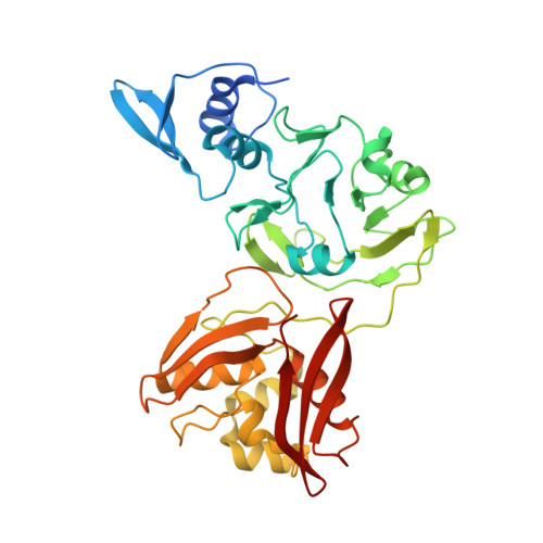
Tipiracil binds to uridine site and inhibits Nsp15 endoribonuclease NendoU from SARS-CoV-2.

Kim, Y.Wower, J.Maltseva, N.Chang, C.Jedrzejczak, R.Wilamowski, M.Kang, S.Nicolaescu, V.Randall, G.Michalska, K.Joachimiak, A.

(2021) Commun Biol 4: 193-193

  • DOI: https://doi.org/10.1038/s42003-021-01735-9
  • Primary Citation of Related Structures:  
    6WLC, 6WXC, 6X1B, 6X4I, 7K1L

  • PubMed Abstract: 
  • SARS-CoV-2 Nsp15 is a uridine-specific endoribonuclease with C-terminal catalytic domain belonging to the EndoU family that is highly conserved in coronaviruses. As endoribonuclease activity seems to be responsible for the interference with the innate immune response, Nsp15 emerges as an attractive target for therapeutic intervention ...

    SARS-CoV-2 Nsp15 is a uridine-specific endoribonuclease with C-terminal catalytic domain belonging to the EndoU family that is highly conserved in coronaviruses. As endoribonuclease activity seems to be responsible for the interference with the innate immune response, Nsp15 emerges as an attractive target for therapeutic intervention. Here we report the first structures with bound nucleotides and show how the enzyme specifically recognizes uridine moiety. In addition to a uridine site we present evidence for a second base binding site that can accommodate any base. The structure with a transition state analog, uridine vanadate, confirms interactions key to catalytic mechanisms. In the presence of manganese ions, the enzyme cleaves unpaired RNAs. This acquired knowledge was instrumental in identifying Tipiracil, an FDA approved drug that is used in the treatment of colorectal cancer, as a potential anti-COVID-19 drug. Using crystallography, biochemical, and whole-cell assays, we demonstrate that Tipiracil inhibits SARS-CoV-2 Nsp15 by interacting with the uridine binding pocket in the enzyme's active site. Our findings provide new insights for the development of uracil scaffold-based drugs.


    Related Citations: 
    • Tipiracil binds to uridine site and inhibits Nsp15 endoribonuclease NendoU from SARS-CoV-2
      Kim, Y., Wower, J., Maltseva, N., Chang, C., Jedrzejczak, R., Wilamowski, M., Kang, S., Nicolaescu,, N., Randall, G., Michalska, K., Joachimiak, A., Center for Structural Genomics of Infectious Diseases (CSGID)
      (2020) Biorxiv --: --

    Organizational Affiliation

    Department of Biochemistry and Molecular Biology, University of Chicago, Chicago, IL, 60367, USA. andrzejj@anl.gov.


Macromolecules
Find similar proteins by:  (by identity cutoff)  |  3D Structure
Entity ID: 1
MoleculeChainsSequence LengthOrganismDetailsImage
Uridylate-specific endoribonuclease
A, B
370Severe acute respiratory syndrome coronavirus 2Mutation(s): 0 
Gene Names: rep1a-1b
EC: 3.1 (PDB Primary Data), 3.4.19.12 (UniProt), 3.4.22 (UniProt), 3.4.22.69 (UniProt), 2.7.7.48 (UniProt), 3.6.4.12 (UniProt), 3.6.4.13 (UniProt), 3.1.13 (UniProt), 4.6.1 (UniProt), 2.1.1.57 (UniProt)
UniProt
Find proteins for P0DTD1 (Severe acute respiratory syndrome coronavirus 2)
Explore P0DTD1 
Go to UniProtKB:  P0DTD1
Entity Groups  
Sequence Clusters30% Identity50% Identity70% Identity90% Identity95% Identity100% Identity
UniProt GroupP0DTD1
Protein Feature View
Expand
  • Reference Sequence
Small Molecules
Ligands 3 Unique
IDChainsName / Formula / InChI Key2D Diagram3D Interactions
U3P (Subject of Investigation/LOI)
Query on U3P

Download Ideal Coordinates CCD File 
C [auth A],
O [auth B]
3'-URIDINEMONOPHOSPHATE
C9 H13 N2 O9 P
FOGRQMPFHUHIGU-XVFCMESISA-N
 Ligand Interaction
EDO
Query on EDO

Download Ideal Coordinates CCD File 
D [auth A]
E [auth A]
F [auth A]
G [auth A]
H [auth A]
D [auth A],
E [auth A],
F [auth A],
G [auth A],
H [auth A],
I [auth A],
J [auth A],
K [auth A],
L [auth A],
M [auth A],
N [auth A],
Q [auth B],
R [auth B],
S [auth B],
T [auth B],
U [auth B],
V [auth B],
W [auth B],
X [auth B],
Y [auth B]
1,2-ETHANEDIOL
C2 H6 O2
LYCAIKOWRPUZTN-UHFFFAOYSA-N
 Ligand Interaction
NA
Query on NA

Download Ideal Coordinates CCD File 
P [auth B]SODIUM ION
Na
FKNQFGJONOIPTF-UHFFFAOYSA-N
 Ligand Interaction
Experimental Data & Validation

Experimental Data

  • Method: X-RAY DIFFRACTION
  • Resolution: 1.85 Å
  • R-Value Free: 0.189 
  • R-Value Work: 0.166 
  • R-Value Observed: 0.167 
  • Space Group: P 63
  • Diffraction Data: https://doi.org/10.18430/m36x4i
Unit Cell:
Length ( Å )Angle ( ˚ )
a = 150.94α = 90
b = 150.94β = 90
c = 111.858γ = 120
Software Package:
Software NamePurpose
PHENIXrefinement
HKL-3000data scaling
SBC-Collectdata collection
PDB_EXTRACTdata extraction
HKL-3000data reduction
HKL-3000phasing

Structure Validation

View Full Validation Report


Ligand Structure Quality Assessment 


Entry History & Funding Information

Deposition Data


Funding OrganizationLocationGrant Number
National Institutes of Health/National Institute Of Allergy and Infectious Diseases (NIH/NIAID)United States--

Revision History  (Full details and data files)

  • Version 1.0: 2020-06-03
    Type: Initial release
  • Version 2.0: 2020-06-10
    Changes: Advisory, Atomic model, Data collection, Database references, Derived calculations, Polymer sequence, Source and taxonomy, Structure summary
  • Version 2.1: 2020-12-09
    Changes: Database references
  • Version 2.2: 2021-02-24
    Changes: Database references