Structural Characterization of Cuta- and Tusavirus: Insight into Protoparvoviruses Capsid Morphology.
Mietzsch, M., McKenna, R., Vaisanen, E., Yu, J.C., Ilyas, M., Hull, J.A., Kurian, J., Smith, J.K., Chipman, P., Lasanajak, Y., Smith, D., Soderlund-Venermo, M., Agbandje-McKenna, M.(2020) Viruses 12
- PubMed: 32560452 Search on PubMedSearch on PubMed Central
- DOI: https://doi.org/10.3390/v12060653
- Primary Citation Related Structures:
6X2I, 6X2K - PubMed Abstract:
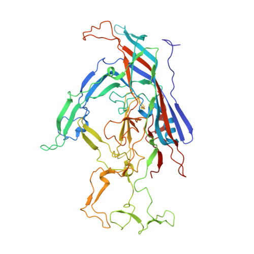
Several members of the Protoparvovirus genus, capable of infecting humans, have been recently discovered, including cutavirus (CuV) and tusavirus (TuV). To begin the characterization of these viruses, we have used cryo-electron microscopy and image reconstruction to determine their capsid structures to ~2.9 Å resolution, and glycan array and cell-based assays to identify glycans utilized for cellular entry. Structural comparisons show that the CuV and TuV capsids share common features with other parvoviruses, including an eight-stranded anti-parallel β-barrel, depressions at the icosahedral 2-fold and surrounding the 5-fold axes, and a channel at the 5-fold axes. However, the viruses exhibit significant topological differences in their viral protein surface loops. These result in three separated 3-fold protrusions, similar to the bufaviruses also infecting humans, suggesting a host-driven structure evolution. The surface loops contain residues involved in receptor binding, cellular trafficking, and antigenic reactivity in other parvoviruses. In addition, terminal sialic acid was identified as the glycan potentially utilized by both CuV and TuV for cellular entry, with TuV showing additional recognition of poly-sialic acid and sialylated Lewis X (sLeXLeXLeX) motifs reported to be upregulated in neurotropic and cancer cells, respectively. These structures provide a platform for annotating the cellular interactions of these human pathogens.
- Department of Biochemistry and Molecular Biology, Center for Structural Biology, McKnight Brain Institute, College of Medicine, University of Florida, Gainesville, FL 32610, USA.
Organizational Affiliation: