6WZB | pdb_00006wzb

Crystal structure of the GltPh V216C-G388C mutant cross-linked with divalent mercury


Experimental Data Snapshot

  • Method: X-RAY DIFFRACTION
  • Resolution: 3.45 Å
  • R-Value Free: 
    0.268 (Depositor), 0.272 (DCC) 
  • R-Value Work: 
    0.233 (Depositor), 0.238 (DCC) 
  • R-Value Observed: 
    0.234 (Depositor) 

Starting Model: experimental
View more details

wwPDB Validation 3D Report Full Report

Validation slider image for 6WZB

This is version 1.3 of the entry. See complete history

Literature

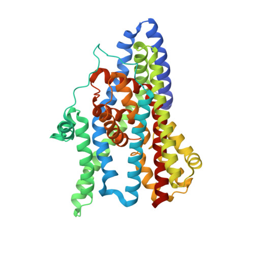
Glutamate transporters have a chloride channel with two hydrophobic gates.

Chen, I.Pant, S.Wu, Q.Cater, R.J.Sobti, M.Vandenberg, R.J.Stewart, A.G.Tajkhorshid, E.Font, J.Ryan, R.M.

(2021) Nature 591: 327-331

  • DOI: https://doi.org/10.1038/s41586-021-03240-9
  • Primary Citation Related Structures: 
    6WYJ, 6WYK, 6WYL, 6WZB, 6X01

  • PubMed Abstract: 

    Glutamate is the most abundant excitatory neurotransmitter in the central nervous system, and its precise control is vital to maintain normal brain function and to prevent excitotoxicity 1 . The removal of extracellular glutamate is achieved by plasma-membrane-bound transporters, which couple glutamate transport to sodium, potassium and pH gradients using an elevator mechanism 2-5 . Glutamate transporters also conduct chloride ions by means of a channel-like process that is thermodynamically uncoupled from transport 6-8 . However, the molecular mechanisms that enable these dual-function transporters to carry out two seemingly contradictory roles are unknown. Here we report the cryo-electron microscopy structure of a glutamate transporter homologue in an open-channel state, which reveals an aqueous cavity that is formed during the glutamate transport cycle. The functional properties of this cavity, combined with molecular dynamics simulations, reveal it to be an aqueous-accessible chloride permeation pathway that is gated by two hydrophobic regions and is conserved across mammalian and archaeal glutamate transporters. Our findings provide insight into the mechanism by which glutamate transporters support their dual function, and add information that will assist in mapping the complete transport cycle shared by the solute carrier 1A transporter family.


  • Organizational Affiliation
    • Transporter Biology Group, School of Medical Sciences, Faculty of Medicine and Health, University of Sydney, Sydney, New South Wales, Australia.

Macromolecule Content 

  • Total Structure Weight: 134.94 kDa 
  • Atom Count: 9,144 
  • Modeled Residue Count: 1,233 
  • Deposited Residue Count: 1,266 
  • Unique protein chains: 1

Macromolecules

Find similar proteins by:|  3D Structure
Entity ID: 1
MoleculeChains  Sequence LengthOrganismDetailsImage
Glutamate transporter homolog
A, B, C
422Pyrococcus horikoshii OT3Mutation(s): 3 
Gene Names: PH1295
Membrane Entity: Yes 
UniProt
Find proteins for O59010 (Pyrococcus horikoshii (strain ATCC 700860 / DSM 12428 / JCM 9974 / NBRC 100139 / OT-3))
Explore O59010 
Go to UniProtKB:  O59010
Entity Groups
Sequence Clusters30% Identity50% Identity70% Identity90% Identity95% Identity100% Identity
UniProt GroupO59010
Sequence Annotations
Expand
Reference Sequence

Small Molecules

Ligands 3 Unique
IDChains Name / Formula / InChI Key2D Diagram3D Interactions
HG

Query on HG


Download:Ideal Coordinates CCD File
G [auth A],
K [auth B],
O [auth C]
MERCURY (II) ION
Hg
BQPIGGFYSBELGY-UHFFFAOYSA-N
ASP

Query on ASP


Download:Ideal Coordinates CCD File
D [auth A],
H [auth B],
L [auth C]
ASPARTIC ACID
C4 H7 N O4
CKLJMWTZIZZHCS-REOHCLBHSA-N
NA

Query on NA


Download:Ideal Coordinates CCD File
E [auth A]
F [auth A]
I [auth B]
J [auth B]
M [auth C]
E [auth A],
F [auth A],
I [auth B],
J [auth B],
M [auth C],
N [auth C]
SODIUM ION
Na
FKNQFGJONOIPTF-UHFFFAOYSA-N

Experimental Data & Validation

Experimental Data

  • Method: X-RAY DIFFRACTION
  • Resolution: 3.45 Å
  • R-Value Free:  0.268 (Depositor), 0.272 (DCC) 
  • R-Value Work:  0.233 (Depositor), 0.238 (DCC) 
  • R-Value Observed: 0.234 (Depositor) 
Space Group: C 2 2 21
Unit Cell:
Length ( Å )Angle ( ˚ )
a = 112.111α = 90
b = 204.573β = 90
c = 207.372γ = 90
Software Package:
Software NamePurpose
PHENIXrefinement
Aimlessdata scaling
PDB_EXTRACTdata extraction
XDSdata reduction
Auto-Rickshawphasing

Structure Validation

View Full Validation Report


Entry History 

& Funding Information

Deposition Data


Funding OrganizationLocationGrant Number
National Health and Medical Research Council (NHMRC, Australia)AustraliaAPP1164494

Revision History  (Full details and data files)

  • Version 1.0: 2021-02-17
    Type: Initial release
  • Version 1.1: 2021-03-03
    Changes: Database references
  • Version 1.2: 2021-03-24
    Changes: Database references
  • Version 1.3: 2023-10-18
    Changes: Data collection, Database references, Refinement description