6WYR | pdb_00006wyr

Crystal structure of anti-Muscle Specific Kinase (MuSK) Fab, MuSK1A

  • Classification: IMMUNE SYSTEM
  • Organism(s): Homo sapiens
  • Expression System: Spodoptera frugiperda
  • Mutation(s): No 

  • Deposited: 2020-05-13 Released: 2020-07-29 
  • Deposition Author(s): Vieni, C., Ekiert, D.
  • Funding Organization(s): National Institutes of Health/National Institute Of Allergy and Infectious Diseases (NIH/NIAID), Muscular Dystrophy Association, Colton Center for Autoimmunity, Brown-Coxe fellowship

Experimental Data Snapshot

  • Method: X-RAY DIFFRACTION
  • Resolution: 1.80 Å
  • R-Value Free: 
    0.213 (Depositor), 0.217 (DCC) 
  • R-Value Work: 
    0.180 (Depositor), 0.185 (DCC) 
  • R-Value Observed: 
    0.181 (Depositor) 

Starting Model: experimental
View more details

wwPDB Validation 3D Report Full Report

Validation slider image for 6WYR

This is version 1.3 of the entry. See complete history

Literature

Affinity maturation is required for pathogenic monovalent IgG4 autoantibody development in myasthenia gravis.

Fichtner, M.L.Vieni, C.Redler, R.L.Kolich, L.Jiang, R.Takata, K.Stathopoulos, P.Suarez, P.A.Nowak, R.J.Burden, S.J.Ekiert, D.C.O'Connor, K.C.

(2020) J Exp Medicine 217

  • DOI: https://doi.org/10.1084/jem.20200513
  • Primary Citation Related Structures: 
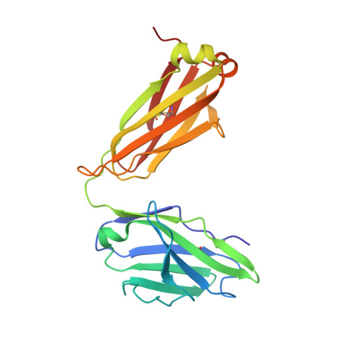
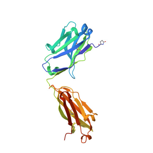
    6WYR, 6WYT

  • PubMed Abstract: 

    Pathogenic muscle-specific tyrosine kinase (MuSK)-specific IgG4 autoantibodies in autoimmune myasthenia gravis (MG) are functionally monovalent as a result of Fab-arm exchange. The development of these unique autoantibodies is not well understood. We examined MG patient-derived monoclonal autoantibodies (mAbs), their corresponding germline-encoded unmutated common ancestors (UCAs), and monovalent antigen-binding fragments (Fabs) to investigate how affinity maturation contributes to binding and immunopathology. Mature mAbs, UCA mAbs, and mature monovalent Fabs bound to MuSK and demonstrated pathogenic capacity. However, monovalent UCA Fabs bound to MuSK but did not have measurable pathogenic capacity. Affinity of the UCA Fabs for MuSK was 100-fold lower than the subnanomolar affinity of the mature Fabs. Crystal structures of two Fabs revealed how mutations acquired during affinity maturation may contribute to increased MuSK-binding affinity. These findings indicate that the autoantigen drives autoimmunity in MuSK MG through the accumulation of somatic mutations such that monovalent IgG4 Fab-arm-exchanged autoantibodies reach a high-affinity threshold required for pathogenic capacity.


  • Organizational Affiliation
    • Department of Neurology, Yale University School of Medicine, New Haven, CT.

Macromolecule Content 

  • Total Structure Weight: 98.21 kDa 
  • Atom Count: 7,496 
  • Modeled Residue Count: 873 
  • Deposited Residue Count: 892 
  • Unique protein chains: 2

Macromolecules

Find similar proteins by:|  3D Structure
Entity ID: 1
MoleculeChains  Sequence LengthOrganismDetailsImage
MuSK1A light chainA [auth L],
C [auth M]
214Homo sapiensMutation(s): 0 
Find similar proteins by:|  3D Structure
Entity ID: 2
MoleculeChains  Sequence LengthOrganismDetailsImage
MuSK1A heavy chainB [auth H],
D [auth I]
232Homo sapiensMutation(s): 0 

Oligosaccharides

Help  
Entity ID: 3
MoleculeChains Length2D Diagram GlycosylationD Interactions
beta-D-mannopyranose-(1-4)-2-acetamido-2-deoxy-beta-D-glucopyranose-(1-4)-2-acetamido-2-deoxy-beta-D-glucopyranoseE [auth A]3N-Glycosylation
Entity ID: 4
MoleculeChains Length2D Diagram GlycosylationD Interactions
2-acetamido-2-deoxy-beta-D-glucopyranose-(1-4)-2-acetamido-2-deoxy-beta-D-glucopyranoseF [auth B]2N-Glycosylation

Small Molecules

Ligands 1 Unique
IDChains Name / Formula / InChI Key2D Diagram3D Interactions
EDO

Query on EDO


Download:Ideal Coordinates CCD File
G [auth L]
H [auth L]
I [auth H]
J [auth H]
K [auth H]
G [auth L],
H [auth L],
I [auth H],
J [auth H],
K [auth H],
L [auth H],
M [auth H],
N [auth M],
O [auth M],
P [auth M],
Q [auth M],
R [auth I],
S [auth I],
T [auth I],
U [auth I],
V [auth I]
1,2-ETHANEDIOL
C2 H6 O2
LYCAIKOWRPUZTN-UHFFFAOYSA-N
Modified Residues  1 Unique
IDChains TypeFormula2D DiagramParent
PCA
Query on PCA
B [auth H],
D [auth I]
L-PEPTIDE LINKINGC5 H7 N O3GLN

Experimental Data & Validation

Experimental Data

  • Method: X-RAY DIFFRACTION
  • Resolution: 1.80 Å
  • R-Value Free:  0.213 (Depositor), 0.217 (DCC) 
  • R-Value Work:  0.180 (Depositor), 0.185 (DCC) 
  • R-Value Observed: 0.181 (Depositor) 
Space Group: P 1 21 1
Unit Cell:
Length ( Å )Angle ( ˚ )
a = 65.87α = 90
b = 71.72β = 104.37
c = 106.18γ = 90
Software Package:
Software NamePurpose
PHENIXrefinement
XDSdata reduction
XSCALEdata scaling
PHASERphasing
PDB_EXTRACTdata extraction
Cootmodel building

Structure Validation

View Full Validation Report


Entry History 

& Funding Information

Deposition Data


Funding OrganizationLocationGrant Number
National Institutes of Health/National Institute Of Allergy and Infectious Diseases (NIH/NIAID)United StatesR01-AI114780
National Institutes of Health/National Institute Of Allergy and Infectious Diseases (NIH/NIAID)United StatesR21-AI142198
Muscular Dystrophy AssociationUnited StatesMDA575198
Colton Center for AutoimmunityUnited States--
Brown-Coxe fellowshipUnited States--

Revision History  (Full details and data files)

  • Version 1.0: 2020-07-29
    Type: Initial release
  • Version 1.1: 2020-09-02
    Changes: Data collection, Database references, Structure summary
  • Version 1.2: 2023-10-18
    Changes: Data collection, Database references, Refinement description
  • Version 1.3: 2024-10-16
    Changes: Structure summary