


Crystal structure of PRL phosphatase C104D mutant in complex with the Bateman domain of CNNM magnesium transporter
Kozlov, G., Funato, Y., Chen, S., Zhang, Z., Illes, K., Miki, H., Gehring, K.(2020) J Biological Chem
Experimental Data Snapshot
Starting Model: experimental
View more details
wwPDB Validation 3D Report Full Report

(2020) J Biological Chem
Entity ID: 1 | |||||
|---|---|---|---|---|---|
| Molecule | Chains | Sequence Length | Organism | Details | Image |
| Metal transporter CNNM2 | 153 | Mus musculus | Mutation(s): 0 Gene Names: Cnnm2, Acdp2 | 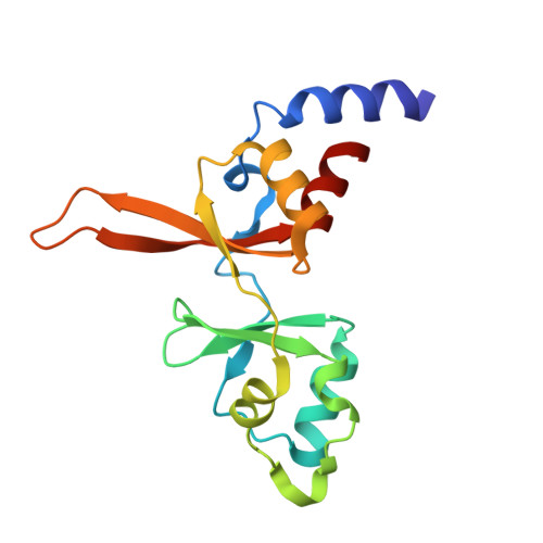 | |
UniProt & NIH Common Fund Data Resources | |||||
IMPC: MGI:2151054 | |||||
Entity Groups | |||||
| Sequence Clusters | 30% Identity50% Identity70% Identity90% Identity95% Identity100% Identity | ||||
| UniProt Group | Q3TWN3 | ||||
Sequence AnnotationsExpand | |||||
Reference Sequence | |||||
Entity ID: 2 | |||||
|---|---|---|---|---|---|
| Molecule | Chains | Sequence Length | Organism | Details | Image |
| Protein tyrosine phosphatase type IVA 1 | 164 | Mus musculus | Mutation(s): 1 Gene Names: Ptp4a1, Prl1 EC: 3.1.3.48 | 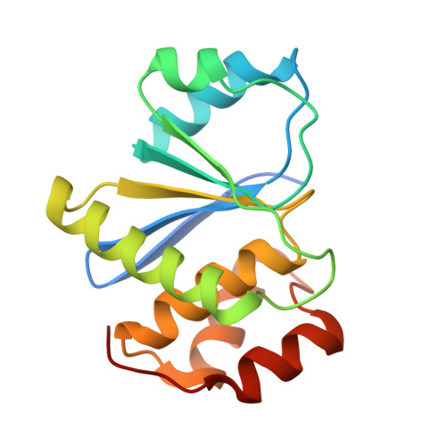 | |
UniProt & NIH Common Fund Data Resources | |||||
IMPC: MGI:1277096 | |||||
Entity Groups | |||||
| Sequence Clusters | 30% Identity50% Identity70% Identity90% Identity95% Identity100% Identity | ||||
| UniProt Group | Q63739 | ||||
Sequence AnnotationsExpand | |||||
Reference Sequence | |||||
| Length ( Å ) | Angle ( ˚ ) |
|---|---|
| a = 52.138 | α = 90 |
| b = 127.935 | β = 90 |
| c = 152.158 | γ = 90 |
| Software Name | Purpose |
|---|---|
| PHENIX | refinement |
| HKL-2000 | data reduction |
| HKL-2000 | data scaling |
| PHASER | phasing |
| Funding Organization | Location | Grant Number |
|---|---|---|
| Natural Sciences and Engineering Research Council (NSERC, Canada) | Canada | RPGIN2014-04686 |


