6WPW

GCGR-Gs signaling complex bound to a designed glucagon derivative


Experimental Data Snapshot

  • Method: ELECTRON MICROSCOPY
  • Resolution: 3.10 Å
  • Aggregation State: PARTICLE 
  • Reconstruction Method: SINGLE PARTICLE 

wwPDB Validation   3D Report Full Report


This is version 1.0 of the entry. See complete history


Literature

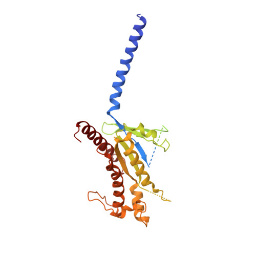
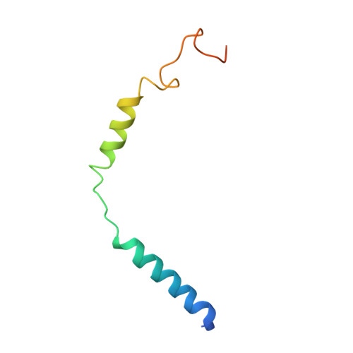
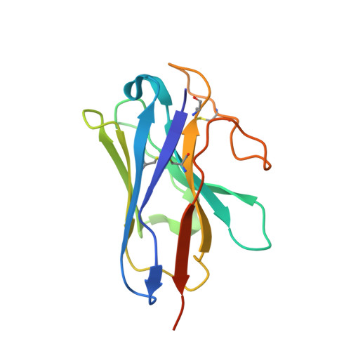
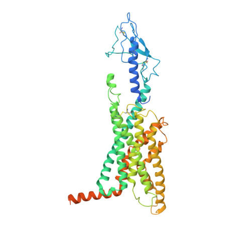
Structural insights into differences in G protein activation by family A and family B GPCRs.

Hilger, D.Kumar, K.K.Hu, H.Pedersen, M.F.O'Brien, E.S.Giehm, L.Jennings, C.Eskici, G.Inoue, A.Lerch, M.Mathiesen, J.M.Skiniotis, G.Kobilka, B.K.

(2020) Science 369

  • DOI: https://doi.org/10.1126/science.aba3373
  • Primary Citation of Related Structures:  
    6WPW

  • PubMed Abstract: 

    Family B heterotrimeric guanine nucleotide-binding protein (G protein)-coupled receptors (GPCRs) play important roles in carbohydrate metabolism. Recent structures of family B GPCR-G s protein complexes reveal a disruption in the α-helix of transmembrane segment 6 (TM6) not observed in family A GPCRs. To investigate the functional impact of this structural difference, we compared the structure and function of the glucagon receptor (GCGR; family B) with the β 2 adrenergic receptor (β 2 AR; family A). We determined the structure of the GCGR-G s complex by means of cryo-electron microscopy at 3.1-angstrom resolution. This structure shows the distinct break in TM6. Guanosine triphosphate (GTP) turnover, guanosine diphosphate release, GTP binding, and G protein dissociation studies revealed much slower rates for G protein activation by the GCGR compared with the β 2 AR. Fluorescence and double electron-electron resonance studies suggest that this difference is due to the inability of agonist alone to induce a detectable outward movement of the cytoplasmic end of TM6.


  • Organizational Affiliation

    Department of Molecular and Cellular Physiology, Stanford University School of Medicine, 279 Campus Drive, Stanford, CA 94305, USA.


Macromolecules
Find similar proteins by:  (by identity cutoff)  |  3D Structure
Entity ID: 1
MoleculeChains Sequence LengthOrganismDetailsImage
Guanine nucleotide-binding protein G(s) subunit alpha isoforms shortA [auth C]380Homo sapiensMutation(s): 0 
Gene Names: GNASGNAS1GSP
Membrane Entity: Yes 
UniProt & NIH Common Fund Data Resources
Find proteins for P63092 (Homo sapiens)
Explore P63092 
Go to UniProtKB:  P63092
PHAROS:  P63092
GTEx:  ENSG00000087460 
Entity Groups  
Sequence Clusters30% Identity50% Identity70% Identity90% Identity95% Identity100% Identity
UniProt GroupP63092
Sequence Annotations
Expand
  • Reference Sequence
Find similar proteins by:  (by identity cutoff)  |  3D Structure
Entity ID: 2
MoleculeChains Sequence LengthOrganismDetailsImage
Guanine nucleotide-binding protein G(I)/G(S)/G(T) subunit beta-1B [auth D]358Homo sapiensMutation(s): 0 
Gene Names: GNB1
UniProt & NIH Common Fund Data Resources
Find proteins for P62873 (Homo sapiens)
Explore P62873 
Go to UniProtKB:  P62873
PHAROS:  P62873
GTEx:  ENSG00000078369 
Entity Groups  
Sequence Clusters30% Identity50% Identity70% Identity90% Identity95% Identity100% Identity
UniProt GroupP62873
Sequence Annotations
Expand
  • Reference Sequence
Find similar proteins by:  (by identity cutoff)  |  3D Structure
Entity ID: 3
MoleculeChains Sequence LengthOrganismDetailsImage
Guanine nucleotide-binding protein G(I)/G(S)/G(O) subunit gamma-2C [auth G]71Homo sapiensMutation(s): 0 
Gene Names: GNG2
Membrane Entity: Yes 
UniProt & NIH Common Fund Data Resources
Find proteins for P59768 (Homo sapiens)
Explore P59768 
Go to UniProtKB:  P59768
PHAROS:  P59768
GTEx:  ENSG00000186469 
Entity Groups  
Sequence Clusters30% Identity50% Identity70% Identity90% Identity95% Identity100% Identity
UniProt GroupP59768
Sequence Annotations
Expand
  • Reference Sequence
Find similar proteins by:  (by identity cutoff)  |  3D Structure
Entity ID: 4
MoleculeChains Sequence LengthOrganismDetailsImage
Nb35D [auth N]138Lama glamaMutation(s): 0 
Entity Groups  
Sequence Clusters30% Identity50% Identity70% Identity90% Identity95% Identity100% Identity
Sequence Annotations
Expand
  • Reference Sequence
Find similar proteins by:  (by identity cutoff)  |  3D Structure
Entity ID: 5
MoleculeChains Sequence LengthOrganismDetailsImage
Glucagon derivative ZP3780E [auth P]29synthetic constructMutation(s): 0 
Entity Groups  
Sequence Clusters30% Identity50% Identity70% Identity90% Identity95% Identity100% Identity
Sequence Annotations
Expand
  • Reference Sequence
Find similar proteins by:  (by identity cutoff)  |  3D Structure
Entity ID: 6
MoleculeChains Sequence LengthOrganismDetailsImage
Glucagon receptorF [auth R]496Homo sapiensMutation(s): 0 
Gene Names: GCGR
Membrane Entity: Yes 
UniProt & NIH Common Fund Data Resources
Find proteins for P47871 (Homo sapiens)
Explore P47871 
Go to UniProtKB:  P47871
PHAROS:  P47871
GTEx:  ENSG00000215644 
Entity Groups  
Sequence Clusters30% Identity50% Identity70% Identity90% Identity95% Identity100% Identity
UniProt GroupP47871
Sequence Annotations
Expand
  • Reference Sequence
Experimental Data & Validation

Experimental Data

  • Method: ELECTRON MICROSCOPY
  • Resolution: 3.10 Å
  • Aggregation State: PARTICLE 
  • Reconstruction Method: SINGLE PARTICLE 
EM Software:
TaskSoftware PackageVersion
RECONSTRUCTIONcisTEM

Structure Validation

View Full Validation Report


Entry History & Funding Information

Deposition Data


Funding OrganizationLocationGrant Number
National Institutes of Health/National Institute of General Medical Sciences (NIH/NIGMS)United StatesR01GM083118
National Institutes of Health/National Institute of General Medical Sciences (NIH/NIGMS)United StatesR01NS028471

Revision History  (Full details and data files)

  • Version 1.0: 2020-08-12
    Type: Initial release