6WMS

Crystal Structure of Human REV-ERBbeta Ligand Binding Domain Co-Bound to Heme and NCoR ID2 Peptide


Experimental Data Snapshot

  • Method: X-RAY DIFFRACTION
  • Resolution: 2.00 Å
  • R-Value Free: 0.239 
  • R-Value Work: 0.188 
  • R-Value Observed: 0.191 

wwPDB Validation   3D Report Full Report


Ligand Structure Quality Assessment 


This is version 1.2 of the entry. See complete history


Literature

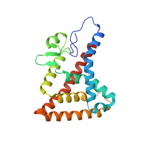
Structural basis for heme-dependent NCoR binding to the transcriptional repressor REV-ERB beta.

Mosure, S.A.Strutzenberg, T.S.Shang, J.Munoz-Tello, P.Solt, L.A.Griffin, P.R.Kojetin, D.J.

(2021) Sci Adv 7

  • DOI: https://doi.org/10.1126/sciadv.abc6479
  • Primary Citation of Related Structures:  
    6WMQ, 6WMS

  • PubMed Abstract: 

    Heme is the endogenous ligand for the constitutively repressive REV-ERB nuclear receptors, REV-ERBα (NR1D1) and REV-ERBβ (NR1D2), but how heme regulates REV-ERB activity remains unclear. Cellular studies indicate that heme is required for the REV-ERBs to bind the corepressor NCoR and repress transcription. However, fluorescence-based biochemical assays suggest that heme displaces NCoR; here, we show that this is due to a heme-dependent artifact. Using ITC and NMR spectroscopy, we show that heme binding remodels the thermodynamic interaction profile of NCoR receptor interaction domain (RID) binding to REV-ERBβ ligand-binding domain (LBD). We solved two crystal structures of REV-ERBβ LBD cobound to heme and NCoR peptides, revealing the heme-dependent NCoR binding mode. ITC and chemical cross-linking mass spectrometry reveals a 2:1 LBD:RID stoichiometry, consistent with cellular studies showing that NCoR-dependent repression of REV-ERB transcription occurs on dimeric DNA response elements. Our findings should facilitate renewed progress toward understanding heme-dependent REV-ERB activity.


  • Organizational Affiliation

    Department of Integrative Structural and Computational Biology, The Scripps Research Institute, Jupiter, FL 33458, USA.


Macromolecules
Find similar proteins by:  (by identity cutoff)  |  3D Structure
Entity ID: 1
MoleculeChains Sequence LengthOrganismDetailsImage
Nuclear receptor Rev-ErbA beta variant 1
A, B
199Homo sapiensMutation(s): 0 
Gene Names: NR1D2
UniProt & NIH Common Fund Data Resources
Find proteins for Q14995 (Homo sapiens)
Explore Q14995 
Go to UniProtKB:  Q14995
PHAROS:  Q14995
GTEx:  ENSG00000174738 
Entity Groups  
Sequence Clusters30% Identity50% Identity70% Identity90% Identity95% Identity100% Identity
UniProt GroupQ14995
Sequence Annotations
Expand
  • Reference Sequence

Find similar proteins by:  Sequence   |   3D Structure  

Entity ID: 2
MoleculeChains Sequence LengthOrganismDetailsImage
NCOR isoform cC [auth E],
D [auth F]
23Homo sapiensMutation(s): 0 
UniProt & NIH Common Fund Data Resources
Find proteins for O75376 (Homo sapiens)
Explore O75376 
Go to UniProtKB:  O75376
PHAROS:  O75376
GTEx:  ENSG00000141027 
Entity Groups  
Sequence Clusters30% Identity50% Identity70% Identity90% Identity95% Identity100% Identity
UniProt GroupO75376
Sequence Annotations
Expand
  • Reference Sequence
Small Molecules
Experimental Data & Validation

Experimental Data

  • Method: X-RAY DIFFRACTION
  • Resolution: 2.00 Å
  • R-Value Free: 0.239 
  • R-Value Work: 0.188 
  • R-Value Observed: 0.191 
  • Space Group: P 1 21 1
Unit Cell:
Length ( Å )Angle ( ˚ )
a = 55.586α = 90
b = 73.4β = 101.91
c = 59.673γ = 90
Software Package:
Software NamePurpose
SCALAdata scaling
PHENIXrefinement
PDB_EXTRACTdata extraction
MOSFLMdata reduction
PHASERphasing

Structure Validation

View Full Validation Report


Ligand Structure Quality Assessment 


Entry History 

Deposition Data

Revision History  (Full details and data files)

  • Version 1.0: 2021-02-17
    Type: Initial release
  • Version 1.1: 2021-02-24
    Changes: Database references
  • Version 1.2: 2023-10-18
    Changes: Data collection, Database references, Refinement description