6WL2 | pdb_00006wl2

preTCRbeta-pMHC complex crystal structure


Experimental Data Snapshot

  • Method: X-RAY DIFFRACTION
  • Resolution: 3.30 Å
  • R-Value Free: 
    0.270 (Depositor), 0.268 (DCC) 
  • R-Value Work: 
    0.228 (Depositor), 0.231 (DCC) 
  • R-Value Observed: 
    0.230 (Depositor) 

Starting Models: experimental
View more details

wwPDB Validation 3D Report Full Report

Validation slider image for 6WL2

This is version 1.4 of the entry. See complete history

Literature

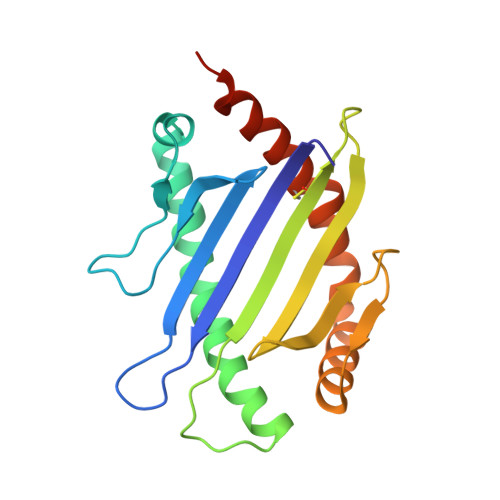
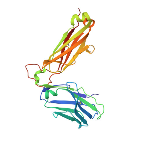
Pre-T cell receptors topologically sample self-ligands during thymocyte beta-selection.

Li, X.Mizsei, R.Tan, K.Mallis, R.J.Duke-Cohan, J.S.Akitsu, A.Tetteh, P.W.Dubey, A.Hwang, W.Wagner, G.Lang, M.J.Arthanari, H.Wang, J.H.Reinherz, E.L.

(2021) Science 371: 181-185

  • DOI: https://doi.org/10.1126/science.abe0918
  • Primary Citation Related Structures: 
    6WL2, 6WL3, 6WL4, 7JI2

  • PubMed Abstract: 

    Self-discrimination, a critical but ill-defined molecular process programmed during thymocyte development, requires myriad pre-T cell receptors (preTCRs) and αβTCRs. Using x-ray crystallography, we show how a preTCR applies the concave β-sheet surface of its single variable domain (Vβ) to "horizontally" grab the protruding MHC α2-helix. By contrast, αβTCRs purpose all six complementarity-determining region (CDR) loops of their paired VαVβ module to recognize peptides bound to major histocompatibility complex molecules (pMHCs) in "vertical" head-to-head binding. The preTCR topological fit ensures that CDR3β reaches the peptide's featured C-terminal segment for pMHC sampling, establishing the subsequent αβTCR canonical docking mode. "Horizontal" docking precludes germline CDR1β- and CDR2β-MHC binding to broaden β-chain repertoire diversification before αβTCR-mediated selection refinement. Thus, one subunit successively attunes the recognition logic of related multicomponent receptors.


  • Organizational Affiliation
    • Laboratory of Immunobiology, Dana-Farber Cancer Institute, Boston, MA, USA. jwang@crystal.harvard.edu ellis_reinherz@dfci.harvard.edu.

Macromolecule Content 

  • Total Structure Weight: 149.18 kDa 
  • Atom Count: 10,279 
  • Modeled Residue Count: 1,265 
  • Deposited Residue Count: 1,296 
  • Unique protein chains: 3

Macromolecules

Find similar proteins by:|  3D Structure
Entity ID: 1
MoleculeChains  Sequence LengthOrganismDetailsImage
H-2 class I histocompatibility antigen, K-B alpha chain
A, D, G
185Mus musculusMutation(s): 2 
Gene Names: H2-K1H2-K
UniProt
Find proteins for P01901 (Mus musculus)
Explore P01901 
Go to UniProtKB:  P01901
Entity Groups
Sequence Clusters30% Identity50% Identity70% Identity90% Identity95% Identity100% Identity
UniProt GroupP01901
Sequence Annotations
Expand
Reference Sequence
Find similar proteins by:  Sequence   |   3D Structure  
Entity ID: 2
MoleculeChains  Sequence LengthOrganismDetailsImage
ARG-GLY-TYR-VAL-TYR-GLN-GLY-LEU
B, E, H
8Vesicular stomatitis virusMutation(s): 0 
UniProt
Find proteins for P03521 (Vesicular stomatitis Indiana virus (strain San Juan))
Explore P03521 
Go to UniProtKB:  P03521
Entity Groups
UniProt GroupP03521
Sequence Annotations
Expand
Reference Sequence
Find similar proteins by:|  3D Structure
Entity ID: 3
MoleculeChains  Sequence LengthOrganismDetailsImage
N15 preTCR beta
C, F, I
239Mus musculusMutation(s): 0 

Experimental Data & Validation

Experimental Data

  • Method: X-RAY DIFFRACTION
  • Resolution: 3.30 Å
  • R-Value Free:  0.270 (Depositor), 0.268 (DCC) 
  • R-Value Work:  0.228 (Depositor), 0.231 (DCC) 
  • R-Value Observed: 0.230 (Depositor) 
Space Group: P 43 21 2
Unit Cell:
Length ( Å )Angle ( ˚ )
a = 108.897α = 90
b = 108.897β = 90
c = 316.173γ = 90
Software Package:
Software NamePurpose
PHENIXrefinement
HKL-3000data reduction
PDB_EXTRACTdata extraction
HKL-3000data scaling
PHENIXphasing

Structure Validation

View Full Validation Report


Entry History 

& Funding Information

Deposition Data


Funding OrganizationLocationGrant Number
National Institutes of Health/National Institute on Drug Abuse (NIH/NIDA)United StatesR01AI136301

Revision History  (Full details and data files)

  • Version 1.0: 2020-12-23
    Type: Initial release
  • Version 1.1: 2020-12-30
    Changes: Database references
  • Version 1.2: 2021-01-20
    Changes: Database references
  • Version 1.3: 2023-10-18
    Changes: Data collection, Database references, Refinement description
  • Version 1.4: 2024-10-30
    Changes: Structure summary