6WIL

CdiB from Acinetobacter baumannii


Experimental Data Snapshot

  • Method: X-RAY DIFFRACTION
  • Resolution: 2.40 Å
  • R-Value Free: 0.257 
  • R-Value Work: 0.208 
  • R-Value Observed: 0.210 

wwPDB Validation   3D Report Full Report


Ligand Structure Quality Assessment 


This is version 1.0 of the entry. See complete history


Literature

Structural insight into toxin secretion by contact dependent growth inhibition transporters.

Guerin, J.Botos, I.Zhang, Z.Lundquist, K.Gumbart, J.C.Buchanan, S.K.

(2020) Elife 9

  • DOI: https://doi.org/10.7554/eLife.58100
  • Primary Citation of Related Structures:  
    6WIL, 6WIM

  • PubMed Abstract: 

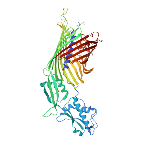
    Bacterial contact-dependent growth inhibition (CDI) systems use a type Vb secretion mechanism to export large CdiA toxins across the outer membrane by dedicated outer membrane transporters called CdiB. Here, we report the first crystal structures of two CdiB transporters from Acinetobacter baumannii and Escherichia coli . CdiB transporters adopt a TpsB fold, containing a 16-stranded transmembrane β-barrel connected to two periplasmic domains. The lumen of the CdiB pore is occluded by an N-terminal α-helix and the conserved extracellular loop 6; these two elements adopt different conformations in the structures. We identified a conserved DxxG motif located on strand β1 that connects loop 6 through different networks of interactions. Structural modifications of DxxG induce rearrangement of extracellular loops and alter interactions with the N-terminal α-helix, preparing the system for α-helix ejection. Using structural biology, functional assays, and molecular dynamics simulations, we show how the barrel pore is primed for CdiA toxin secretion.


  • Organizational Affiliation

    Laboratory of Molecular Biology, NIDDK, NIH, Bethesda, United States.


Macromolecules
Find similar proteins by:  (by identity cutoff)  |  3D Structure
Entity ID: 1
MoleculeChains Sequence LengthOrganismDetailsImage
Hemolysin activator protein CdiB560Acinetobacter baumannii ACICUMutation(s): 0 
Gene Names: ACICU_01912
Membrane Entity: Yes 
UniProt
Find proteins for A0A241YIJ9 (Acinetobacter baumannii)
Explore A0A241YIJ9 
Go to UniProtKB:  A0A241YIJ9
Entity Groups  
Sequence Clusters30% Identity50% Identity70% Identity90% Identity95% Identity100% Identity
UniProt GroupA0A241YIJ9
Sequence Annotations
Expand
  • Reference Sequence
Experimental Data & Validation

Experimental Data

  • Method: X-RAY DIFFRACTION
  • Resolution: 2.40 Å
  • R-Value Free: 0.257 
  • R-Value Work: 0.208 
  • R-Value Observed: 0.210 
  • Space Group: P 1
Unit Cell:
Length ( Å )Angle ( ˚ )
a = 46.951α = 100.8
b = 49.283β = 90.37
c = 86.884γ = 109.94
Software Package:
Software NamePurpose
PHENIXrefinement
PDB_EXTRACTdata extraction
XDSdata reduction
XSCALEdata scaling
PHENIXphasing

Structure Validation

View Full Validation Report


Ligand Structure Quality Assessment 


Entry History & Funding Information

Deposition Data


Funding OrganizationLocationGrant Number
National Science Foundation (United States)United StatesOCI-1053575

Revision History  (Full details and data files)

  • Version 1.0: 2020-11-04
    Type: Initial release