6WII | pdb_00006wii

Crystal structure of the K. pneumoniae LpxH/JH-LPH-41 complex


Experimental Data Snapshot

  • Method: X-RAY DIFFRACTION
  • Resolution: 1.85 Å
  • R-Value Free: 
    0.187 (Depositor), 0.188 (DCC) 
  • R-Value Work: 
    0.160 (Depositor), 0.162 (DCC) 
  • R-Value Observed: 
    0.162 (Depositor) 

Starting Model: experimental
View more details

wwPDB Validation 3D Report Full Report

Validation slider image for 6WII

Ligand Structure Quality Assessment 


This is version 1.1 of the entry. See complete history

Literature

Synthesis and evaluation of sulfonyl piperazine LpxH inhibitors.

Kwak, S.H.Cochrane, C.S.Ennis, A.F.Lim, W.Y.Webster, C.G.Cho, J.Fenton, B.A.Zhou, P.Hong, J.

(2020) Bioorg Chem 102: 104055-104055

  • DOI: https://doi.org/10.1016/j.bioorg.2020.104055
  • Primary Citation Related Structures: 
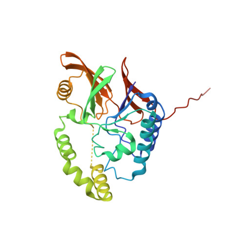
    6WII

  • PubMed Abstract: 

    The UDP-2,3-diacylglucosamine pyrophosphate hydrolase LpxH is essential in lipid A biosynthesis and has emerged as a promising target for the development of novel antibiotics against multidrug-resistant Gram-negative pathogens. Recently, we reported the crystal structure of Klebsiella pneumoniae LpxH in complex with 1 (AZ1), a sulfonyl piperazine LpxH inhibitor. The analysis of the LpxH-AZ1 co-crystal structure and ligand dynamics led to the design of 2 (JH-LPH-28) and 3 (JH-LPH-33) with enhanced LpxH inhibition. In order to harness our recent findings, we prepared and evaluated a series of sulfonyl piperazine analogs with modifications in the phenyl and N-acetyl groups of 3. Herein, we describe the synthesis and structure-activity relationship of sulfonyl piperazine LpxH inhibitors. We also report the structural analysis of an extended N-acyl chain analog 27b (JH-LPH-41) in complex with K. pneumoniae LpxH, revealing that 27b reaches an untapped polar pocket near the di-manganese cluster in the active site of K. pneumoniae LpxH. We expect that our findings will provide designing principles for new LpxH inhibitors and establish important frameworks for the future development of antibiotics against multidrug-resistant Gram-negative pathogens.


  • Organizational Affiliation
    • Department of Chemistry, Duke University, Durham, NC 27708, United States.

Macromolecule Content 

  • Total Structure Weight: 30.64 kDa 
  • Atom Count: 2,171 
  • Modeled Residue Count: 238 
  • Deposited Residue Count: 259 
  • Unique protein chains: 1

Macromolecules

Find similar proteins by:|  3D Structure
Entity ID: 1
MoleculeChains  Sequence LengthOrganismDetailsImage
UDP-2,3-diacylglucosamine hydrolase259Klebsiella pneumoniaeMutation(s): 0 
Gene Names: lpxH_1lpxHC3483_19950NCTC9128_00880SAMEA104305404_03891
EC: 3.6.1.54
Membrane Entity: Yes 
UniProt
Find proteins for A6T5R0 (Klebsiella pneumoniae subsp. pneumoniae (strain ATCC 700721 / MGH 78578))
Explore A6T5R0 
Go to UniProtKB:  A6T5R0
Entity Groups
Sequence Clusters30% Identity50% Identity70% Identity90% Identity95% Identity100% Identity
UniProt GroupA6T5R0
Sequence Annotations
Expand
Reference Sequence

Small Molecules

Ligands 4 Unique
IDChains Name / Formula / InChI Key2D Diagram3D Interactions
U2V
(Subject of Investigation/LOI)

Query on U2V


Download:Ideal Coordinates CCD File
B [auth A]5-({[4-({4-[3-chloro-5-(trifluoromethyl)phenyl]piperazin-1-yl}sulfonyl)phenyl]carbamoyl}amino)-N-hydroxypentanamide
C23 H27 Cl F3 N5 O5 S
YNBLMVJKYWHKRJ-UHFFFAOYSA-N
GOL

Query on GOL


Download:Ideal Coordinates CCD File
H [auth A]GLYCEROL
C3 H8 O3
PEDCQBHIVMGVHV-UHFFFAOYSA-N
DMS

Query on DMS


Download:Ideal Coordinates CCD File
E [auth A],
F [auth A],
G [auth A]
DIMETHYL SULFOXIDE
C2 H6 O S
IAZDPXIOMUYVGZ-UHFFFAOYSA-N
MN

Query on MN


Download:Ideal Coordinates CCD File
C [auth A],
D [auth A]
MANGANESE (II) ION
Mn
WAEMQWOKJMHJLA-UHFFFAOYSA-N
Binding Affinity Annotations 
IDSourceBinding Affinity
U2V BindingDB:  6WII IC50: 73 (nM) from 1 assay(s)

Experimental Data & Validation

Experimental Data

  • Method: X-RAY DIFFRACTION
  • Resolution: 1.85 Å
  • R-Value Free:  0.187 (Depositor), 0.188 (DCC) 
  • R-Value Work:  0.160 (Depositor), 0.162 (DCC) 
  • R-Value Observed: 0.162 (Depositor) 
Space Group: P 32 2 1
Unit Cell:
Length ( Å )Angle ( ˚ )
a = 105.71α = 90
b = 105.71β = 90
c = 53.65γ = 120
Software Package:
Software NamePurpose
XDSdata reduction
XSCALEdata scaling
PHASERphasing
PHENIXrefinement

Structure Validation

View Full Validation Report


Ligand Structure Quality Assessment 


Entry History 

& Funding Information

Deposition Data


Funding OrganizationLocationGrant Number
National Institutes of Health/National Institute Of Allergy and Infectious Diseases (NIH/NIAID)United StatesAI139216

Revision History  (Full details and data files)

  • Version 1.0: 2020-07-29
    Type: Initial release
  • Version 1.1: 2023-10-18
    Changes: Data collection, Database references, Refinement description