6WIG

Structure of STENOFOLIA Protein HD domain bound with DNA


Experimental Data Snapshot

  • Method: X-RAY DIFFRACTION
  • Resolution: 2.10 Å
  • R-Value Free: 0.250 
  • R-Value Work: 0.194 
  • R-Value Observed: 0.200 

wwPDB Validation   3D Report Full Report


This is version 1.1 of the entry. See complete history


Literature

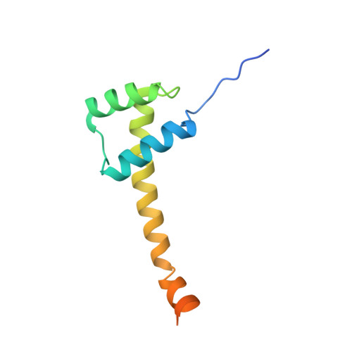
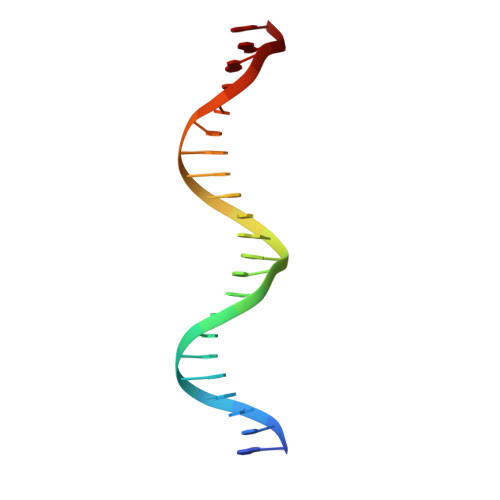
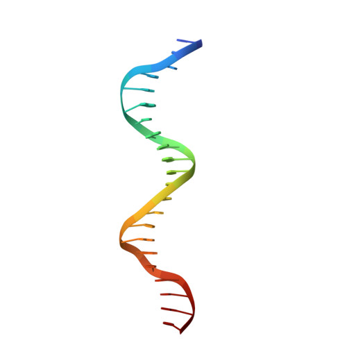
Structure of the unique tetrameric STENOFOLIA homeodomain bound with target promoter DNA.

Pathak, P.K.Zhang, F.Peng, S.Niu, L.Chaturvedi, J.Elliott, J.Xiang, Y.Tadege, M.Deng, J.

(2021) Acta Crystallogr D Struct Biol 77: 1050-1063

  • DOI: https://doi.org/10.1107/S205979832100632X
  • Primary Citation of Related Structures:  
    6WIG

  • PubMed Abstract: 

    Homeobox transcription factors are key regulators of morphogenesis and development in both animals and plants. In plants, the WUSCHEL-related homeobox (WOX) family of transcription factors function as central organizers of several developmental programs ranging from embryo patterning to meristematic stem-cell maintenance through transcriptional activation and repression mechanisms. The Medicago truncatula STENOFOLIA (STF) gene is a master regulator of leaf-blade lateral development. Here, the crystal structure of the homeodomain (HD) of STF (STF-HD) in complex with its promoter DNA is reported at 2.1 Å resolution. STF-HD binds DNA as a tetramer, enclosing nearly the entire bound DNA surface. The STF-HD tetramer is partially stabilized by docking of the C-terminal tail of one protomer onto a conserved hydrophobic surface on the head of another protomer in a head-to-tail manner. STF-HD specifically binds TGA motifs, although the promoter sequence also contains TAAT motifs. Helix α3 not only serves a canonical role as a base reader in the major groove, but also provides DNA binding in the minor groove through basic residues located at its C-terminus. The structural and functional data in planta reported here provide new insights into the DNA-binding mechanisms of plant-specific HDs from the WOX family of transcription factors.


  • Organizational Affiliation

    Department of Biochemistry and Molecular Biology, Oklahoma State University, Stillwater, OK 74078, USA.


Macromolecules

Find similar proteins by:  (by identity cutoff)  |  3D Structure
Entity ID: 1
MoleculeChains Sequence LengthOrganismDetailsImage
STENOFOLIA
A, B
106Medicago truncatulaMutation(s): 0 
Gene Names: STF
UniProt
Find proteins for G0ZGT3 (Medicago truncatula)
Explore G0ZGT3 
Go to UniProtKB:  G0ZGT3
Entity Groups  
Sequence Clusters30% Identity50% Identity70% Identity90% Identity95% Identity100% Identity
UniProt GroupG0ZGT3
Sequence Annotations
Expand
  • Reference Sequence

Find similar nucleic acids by:  Sequence   |   3D Structure  

Entity ID: 2
MoleculeChains LengthOrganismImage
DNA (5'-D(P*GP*CP*AP*AP*AP*TP*TP*AP*AP*TP*GP*AP*TP*TP*TP*AP*TP*TP*CP*AP*AP*G)-3')22Medicago truncatula
Sequence Annotations
Expand
  • Reference Sequence

Find similar nucleic acids by:  Sequence   |   3D Structure  

Entity ID: 3
MoleculeChains LengthOrganismImage
DNA (5'-D(P*CP*TP*TP*GP*AP*AP*TP*AP*AP*AP*TP*CP*AP*TP*TP*AP*AP*TP*TP*TP*GP*C)-3')22Medicago truncatula
Sequence Annotations
Expand
  • Reference Sequence
Experimental Data & Validation

Experimental Data

  • Method: X-RAY DIFFRACTION
  • Resolution: 2.10 Å
  • R-Value Free: 0.250 
  • R-Value Work: 0.194 
  • R-Value Observed: 0.200 
  • Space Group: P 1 21 1
Unit Cell:
Length ( Å )Angle ( ˚ )
a = 48.061α = 90
b = 49.489β = 106.46
c = 69.81γ = 90
Software Package:
Software NamePurpose
PHENIXrefinement
PDB_EXTRACTdata extraction
HKL-3000data reduction
HKL-3000data scaling
PHENIXphasing

Structure Validation

View Full Validation Report


Entry History 

Deposition Data

Revision History  (Full details and data files)

  • Version 1.0: 2021-08-11
    Type: Initial release
  • Version 1.1: 2022-02-23
    Changes: Database references