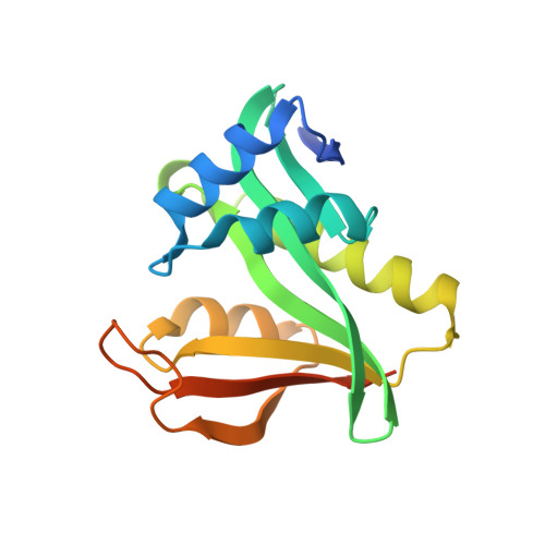
6WFG

Crystal structure of human Naa50 in complex with an inhibitor (compound 3) identified using DNA encoded library technology


Experimental Data Snapshot

  • Method: X-RAY DIFFRACTION
  • Resolution: 2.16 Å
  • R-Value Free: 0.253 
  • R-Value Work: 0.201 
  • R-Value Observed: 0.203 

wwPDB Validation   3D Report Full Report


This is version 1.0 of the entry. See complete history


Literature

Characterization of SpecificN-alpha-Acetyltransferase 50 (Naa50) Inhibitors Identified Using a DNA Encoded Library.

Kung, P.P.Bingham, P.Burke, B.J.Chen, Q.Cheng, X.Deng, Y.L.Dou, D.Feng, J.Gallego, G.M.Gehring, M.R.Grant, S.K.Greasley, S.Harris, A.R.Maegley, K.A.Meier, J.Meng, X.Montano, J.L.Morgan, B.A.Naughton, B.S.Palde, P.B.Paul, T.A.Richardson, P.Sakata, S.Shaginian, A.Sonnenburg, W.K.Subramanyam, C.Timofeevski, S.Wan, J.Yan, W.Stewart, A.E.

(2020) ACS Med Chem Lett 11: 1175-1184

  • DOI: https://doi.org/10.1021/acsmedchemlett.0c00029
  • Primary Citation of Related Structures:  
    6WF3, 6WF5, 6WFG, 6WFK, 6WFN, 6WFO

  • PubMed Abstract: 

    Two novel compounds were identified as Naa50 binders/inhibitors using DNA-encoded technology screening. Biophysical and biochemical data as well as cocrystal structures were obtained for both compounds ( 3a and 4a ) to understand their mechanism of action. These data were also used to rationalize the binding affinity differences observed between the two compounds and a MLGP peptide-containing substrate. Cellular target engagement experiments further confirm the Naa50 binding of 4a and demonstrate its selectivity toward related enzymes (Naa10 and Naa60). Additional analogs of inhibitor 4a were also evaluated to study the binding mode observed in the cocrystal structures.


  • Organizational Affiliation

    Chemical Biology Laboratory, National Cancer Institute, Frederick, Maryland 21702, United States.


Macromolecules
Find similar proteins by:  (by identity cutoff)  |  3D Structure
Entity ID: 1
MoleculeChains Sequence LengthOrganismDetailsImage
N-alpha-acetyltransferase 50A,
B [auth C],
C [auth E]
171Homo sapiensMutation(s): 0 
Gene Names: NAA50MAK3NAT13NAT5
EC: 2.3.1.258 (PDB Primary Data), 2.3.1 (PDB Primary Data)
UniProt & NIH Common Fund Data Resources
Find proteins for Q9GZZ1 (Homo sapiens)
Explore Q9GZZ1 
Go to UniProtKB:  Q9GZZ1
PHAROS:  Q9GZZ1
Entity Groups  
Sequence Clusters30% Identity50% Identity70% Identity90% Identity95% Identity100% Identity
UniProt GroupQ9GZZ1
Sequence Annotations
Expand
  • Reference Sequence
Small Molecules
Ligands 2 Unique
IDChains Name / Formula / InChI Key2D Diagram3D Interactions
COA
Query on COA

Download Ideal Coordinates CCD File 
D [auth A],
F [auth C],
H [auth E]
COENZYME A
C21 H36 N7 O16 P3 S
RGJOEKWQDUBAIZ-IBOSZNHHSA-N
U3V (Subject of Investigation/LOI)
Query on U3V

Download Ideal Coordinates CCD File 
E [auth A],
G [auth C],
I [auth E]
(2S)-N-[(2S)-3-[1-(3-tert-butyl-1-methyl-1H-pyrazole-5-carbonyl)piperidin-4-yl]-1-(methylamino)-1-oxopropan-2-yl]-6-oxopiperidine-2-carboxamide
C24 H38 N6 O4
SLKUTPSFHQUWLD-IRXDYDNUSA-N
Binding Affinity Annotations 
IDSourceBinding Affinity
U3V Binding MOAD:  6WFG IC50: 2700 (nM) from 1 assay(s)
Experimental Data & Validation

Experimental Data

  • Method: X-RAY DIFFRACTION
  • Resolution: 2.16 Å
  • R-Value Free: 0.253 
  • R-Value Work: 0.201 
  • R-Value Observed: 0.203 
  • Space Group: P 1 21 1
Unit Cell:
Length ( Å )Angle ( ˚ )
a = 47.83α = 90
b = 102.94β = 106.71
c = 67.64γ = 90
Software Package:
Software NamePurpose
XDSdata reduction
Aimlessdata scaling
BUSTERrefinement
PDB_EXTRACTdata extraction
autoBUSTERphasing

Structure Validation

View Full Validation Report


Entry History 

Deposition Data

Revision History  (Full details and data files)

  • Version 1.0: 2020-07-01
    Type: Initial release