6WDV

Crystal Structure of Danio rerio Histone Deacetylase 10 in Complex with Dimethylaminomethylindole Phenylhydroxamate Inhibitor


Experimental Data Snapshot

  • Method: X-RAY DIFFRACTION
  • Resolution: 2.40 Å
  • R-Value Free: 0.227 
  • R-Value Work: 0.181 
  • R-Value Observed: 0.184 

wwPDB Validation   3D Report Full Report


Ligand Structure Quality Assessment 


This is version 1.3 of the entry. See complete history


Literature

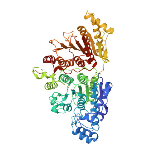
Structural Basis for the Selective Inhibition of HDAC10, the Cytosolic Polyamine Deacetylase.

Herbst-Gervasoni, C.J.Steimbach, R.R.Morgen, M.Miller, A.K.Christianson, D.W.

(2020) ACS Chem Biol 15: 2154-2163

  • DOI: https://doi.org/10.1021/acschembio.0c00362
  • Primary Citation of Related Structures:  
    6WBQ, 6WDV, 6WDW, 6WDX, 6WDY

  • PubMed Abstract: 

    The cytosolic class IIb histone deacetylase HDAC10 is an emerging target for drug design. As an inducer of autophagy, its selective inhibition suppresses the autophagic response that otherwise attenuates the efficacy of cytotoxic cancer chemotherapy drugs. HDAC10 is a zinc-dependent polyamine deacetylase exhibiting maximal catalytic activity against N 8 -acetylspermidine. As revealed in the structure of Danio rerio (zebrafish) HDAC10, two conserved structural motifs direct this narrow substrate specificity: a 3 10 helix containing the P(E,A)CE motif that sterically constricts the active site and an electrostatic "gatekeeper," E274, that confers selectivity for cationic polyamine substrates. To accelerate drug design efforts targeting human HDAC10, we now report the preparation of "humanized" zebrafish HDAC10 in which two amino acid substitutions, A24E and D94A, yield an active site contour more similar to that of human HDAC10. X-ray crystal structures of this HDAC10 variant complexed with Tubastatin A and indole analogues bearing pendant tertiary amines reveal that inhibitors capable of hydrogen bonding with gatekeeper E274 exhibit high affinity and selectivity for HDAC10 over HDAC6 (the other class IIb isozyme). Moreover, these structures reveal that the P(E,A)CE motif helix can shift by up to 2 Å to accommodate the binding of bulky inhibitors. Thus, slender polyamine-like inhibitor structures are not exclusively required for selective, high affinity binding to HDAC10. Indeed, the flexibility of the P(E,A)CE motif helix could conceivably enable the binding of certain protein substrates.


  • Organizational Affiliation

    Roy and Diana Vagelos Laboratories, Department of Chemistry, University of Pennsylvania, Philadelphia, Pennsylvania 19104-6323, United States.


Macromolecules
Find similar proteins by:  (by identity cutoff)  |  3D Structure
Entity ID: 1
MoleculeChains Sequence LengthOrganismDetailsImage
Polyamine deacetylase HDAC10676Danio rerioMutation(s): 0 
Gene Names: hdac10
EC: 3.5.1.48 (PDB Primary Data), 3.5.1.62 (PDB Primary Data)
UniProt
Find proteins for F1QCV2 (Danio rerio)
Explore F1QCV2 
Go to UniProtKB:  F1QCV2
Entity Groups  
Sequence Clusters30% Identity50% Identity70% Identity90% Identity95% Identity100% Identity
UniProt GroupF1QCV2
Sequence Annotations
Expand
  • Reference Sequence
Small Molecules
Ligands 4 Unique
IDChains Name / Formula / InChI Key2D Diagram3D Interactions
TWM (Subject of Investigation/LOI)
Query on TWM

Download Ideal Coordinates CCD File 
B [auth A]4-({3-[(dimethylamino)methyl]-1H-indol-1-yl}methyl)-N-hydroxybenzamide
C19 H21 N3 O2
LYCCCRFBRDLGGA-UHFFFAOYSA-N
PO4
Query on PO4

Download Ideal Coordinates CCD File 
C [auth A],
D [auth A]
PHOSPHATE ION
O4 P
NBIIXXVUZAFLBC-UHFFFAOYSA-K
ZN
Query on ZN

Download Ideal Coordinates CCD File 
E [auth A]ZINC ION
Zn
PTFCDOFLOPIGGS-UHFFFAOYSA-N
K
Query on K

Download Ideal Coordinates CCD File 
F [auth A],
G [auth A]
POTASSIUM ION
K
NPYPAHLBTDXSSS-UHFFFAOYSA-N
Binding Affinity Annotations 
IDSourceBinding Affinity
TWM Binding MOAD:  6WDV IC50: 5 (nM) from 1 assay(s)
Experimental Data & Validation

Experimental Data

  • Method: X-RAY DIFFRACTION
  • Resolution: 2.40 Å
  • R-Value Free: 0.227 
  • R-Value Work: 0.181 
  • R-Value Observed: 0.184 
  • Space Group: P 31 2 1
Unit Cell:
Length ( Å )Angle ( ˚ )
a = 80.67α = 90
b = 80.67β = 90
c = 246.842γ = 120
Software Package:
Software NamePurpose
PHENIXrefinement
iMOSFLMdata reduction
Aimlessdata scaling
PHASERphasing

Structure Validation

View Full Validation Report


Ligand Structure Quality Assessment 


Entry History & Funding Information

Deposition Data


Funding OrganizationLocationGrant Number
National Institutes of Health/National Human Genome Research Institute (NIH/NHGRI)United StatesGM49758

Revision History  (Full details and data files)

  • Version 1.0: 2020-07-22
    Type: Initial release
  • Version 1.1: 2020-07-29
    Changes: Database references, Derived calculations
  • Version 1.2: 2020-09-02
    Changes: Database references
  • Version 1.3: 2023-10-18
    Changes: Data collection, Database references, Refinement description