6WBM

Cryo-EM structure of human Pannexin 1 channel N255A mutant

  • Classification: TRANSPORT PROTEIN
  • Organism(s): Homo sapiens
  • Expression System: Homo sapiens
  • Mutation(s): Yes 
  • Membrane Protein: Yes  PDBTMMemProtMDmpstruc

  • Deposited: 2020-03-26 Released: 2020-06-03 
  • Deposition Author(s): Lu, W., Du, J., Ruan, Z.
  • Funding Organization(s): National Institutes of Health/National Heart, Lung, and Blood Institute (NIH/NHLBI), National Institutes of Health/National Institute of Neurological Disorders and Stroke (NIH/NINDS), American Heart Association

Experimental Data Snapshot

  • Method: ELECTRON MICROSCOPY
  • Resolution: 2.86 Å
  • Aggregation State: PARTICLE 
  • Reconstruction Method: SINGLE PARTICLE 

wwPDB Validation   3D Report Full Report


This is version 2.0 of the entry. See complete history


Literature

Structures of human pannexin 1 reveal ion pathways and mechanism of gating.

Ruan, Z.Orozco, I.J.Du, J.Lu, W.

(2020) Nature 584: 646-651

  • DOI: https://doi.org/10.1038/s41586-020-2357-y
  • Primary Citation of Related Structures:  
    6WBF, 6WBG, 6WBI, 6WBK, 6WBL, 6WBM, 6WBN

  • PubMed Abstract: 

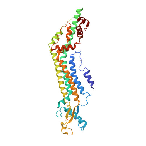
    Pannexin 1 (PANX1) is an ATP-permeable channel with critical roles in a variety of physiological functions such as blood pressure regulation 1 , apoptotic cell clearance 2 and human oocyte development 3 . Here we present several structures of human PANX1 in a heptameric assembly at resolutions of up to 2.8 angström, including an apo state, a caspase-7-cleaved state and a carbenoxolone-bound state. We reveal a gating mechanism that involves two ion-conducting pathways. Under normal cellular conditions, the intracellular entry of the wide main pore is physically plugged by the C-terminal tail. Small anions are conducted through narrow tunnels in the intracellular domain. These tunnels connect to the main pore and are gated by a long linker between the N-terminal helix and the first transmembrane helix. During apoptosis, the C-terminal tail is cleaved by caspase, allowing the release of ATP through the main pore. We identified a carbenoxolone-binding site embraced by W74 in the extracellular entrance and a role for carbenoxolone as a channel blocker. We identified a gap-junction-like structure using a glycosylation-deficient mutant, N255A. Our studies provide a solid foundation for understanding the molecular mechanisms underlying the channel gating and inhibition of PANX1 and related large-pore channels.


  • Organizational Affiliation

    Van Andel Institute, Grand Rapids, MI, USA.


Macromolecules
Find similar proteins by:  (by identity cutoff)  |  3D Structure
Entity ID: 1
MoleculeChains Sequence LengthOrganismDetailsImage
Pannexin-1
A, B, C, D, E
A, B, C, D, E, F, G
373Homo sapiensMutation(s): 1 
Gene Names: PANX1MRS1UNQ2529/PRO6028
Membrane Entity: Yes 
UniProt & NIH Common Fund Data Resources
Find proteins for Q96RD7 (Homo sapiens)
Explore Q96RD7 
Go to UniProtKB:  Q96RD7
PHAROS:  Q96RD7
GTEx:  ENSG00000110218 
Entity Groups  
Sequence Clusters30% Identity50% Identity70% Identity90% Identity95% Identity100% Identity
UniProt GroupQ96RD7
Sequence Annotations
Expand
  • Reference Sequence
Small Molecules
Ligands 4 Unique
IDChains Name / Formula / InChI Key2D Diagram3D Interactions
3PE
Query on 3PE

Download Ideal Coordinates CCD File 
CA [auth D]
IA [auth E]
J [auth A]
OA [auth F]
Q [auth B]
CA [auth D],
IA [auth E],
J [auth A],
OA [auth F],
Q [auth B],
UA [auth G],
W [auth C]
1,2-Distearoyl-sn-glycerophosphoethanolamine
C41 H82 N O8 P
LVNGJLRDBYCPGB-LDLOPFEMSA-N
PTY
Query on PTY

Download Ideal Coordinates CCD File 
AA [auth D]
BA [auth D]
GA [auth E]
H [auth A]
HA [auth E]
AA [auth D],
BA [auth D],
GA [auth E],
H [auth A],
HA [auth E],
I [auth A],
MA [auth F],
NA [auth F],
O [auth B],
P [auth B],
SA [auth G],
TA [auth G],
U [auth C],
V [auth C]
PHOSPHATIDYLETHANOLAMINE
C40 H80 N O8 P
NJGIRBISCGPRPF-KXQOOQHDSA-N
DGA
Query on DGA

Download Ideal Coordinates CCD File 
FA [auth D]
K [auth A]
LA [auth E]
N [auth A]
RA [auth F]
FA [auth D],
K [auth A],
LA [auth E],
N [auth A],
RA [auth F],
T [auth B],
Z [auth C]
DIACYL GLYCEROL
C39 H76 O5
UHUSDOQQWJGJQS-QNGWXLTQSA-N
CLR
Query on CLR

Download Ideal Coordinates CCD File 
DA [auth D]
EA [auth D]
JA [auth E]
KA [auth E]
L [auth A]
DA [auth D],
EA [auth D],
JA [auth E],
KA [auth E],
L [auth A],
M [auth A],
PA [auth F],
QA [auth F],
R [auth B],
S [auth B],
VA [auth G],
WA [auth G],
X [auth C],
Y [auth C]
CHOLESTEROL
C27 H46 O
HVYWMOMLDIMFJA-DPAQBDIFSA-N
Experimental Data & Validation

Experimental Data

  • Method: ELECTRON MICROSCOPY
  • Resolution: 2.86 Å
  • Aggregation State: PARTICLE 
  • Reconstruction Method: SINGLE PARTICLE 
EM Software:
TaskSoftware PackageVersion
MODEL REFINEMENTPHENIX
RECONSTRUCTIONRELION3.1b

Structure Validation

View Full Validation Report


Entry History & Funding Information

Deposition Data


Funding OrganizationLocationGrant Number
National Institutes of Health/National Heart, Lung, and Blood Institute (NIH/NHLBI)United StatesR56HL144929
National Institutes of Health/National Institute of Neurological Disorders and Stroke (NIH/NINDS)United StatesR01NS111031
American Heart AssociationUnited States20POST35120556

Revision History  (Full details and data files)

  • Version 1.0: 2020-06-03
    Type: Initial release
  • Version 1.1: 2020-06-17
    Changes: Database references
  • Version 1.2: 2020-09-09
    Changes: Database references
  • Version 2.0: 2021-06-30
    Changes: Advisory, Atomic model, Data collection, Derived calculations, Non-polymer description, Structure summary