6W7V | pdb_00006w7v

Structure of rabbit actin in complex with truncated analog of Mycalolide B


Experimental Data Snapshot

  • Method: X-RAY DIFFRACTION
  • Resolution: 1.70 Å
  • R-Value Free: 
    0.213 (Depositor), 0.210 (DCC) 
  • R-Value Work: 
    0.179 (Depositor), 0.174 (DCC) 
  • R-Value Observed: 
    0.181 (Depositor) 

Starting Model: experimental
View more details

wwPDB Validation 3D Report Full Report

Validation slider image for 6W7V

Ligand Structure Quality Assessment 


This is version 1.3 of the entry. See complete history

Literature

Truncated Actin-Targeting Macrolide Derivative Blocks Cancer Cell Motility and Invasion of Extracellular Matrix.

Pipaliya, B.V.Trofimova, D.N.Grange, R.L.Aeluri, M.Deng, X.Shah, K.Craig, A.W.Allingham, J.S.Evans, P.A.

(2021) J Am Chem Soc 143: 6847-6854

  • DOI: https://doi.org/10.1021/jacs.0c12404
  • Primary Citation Related Structures: 
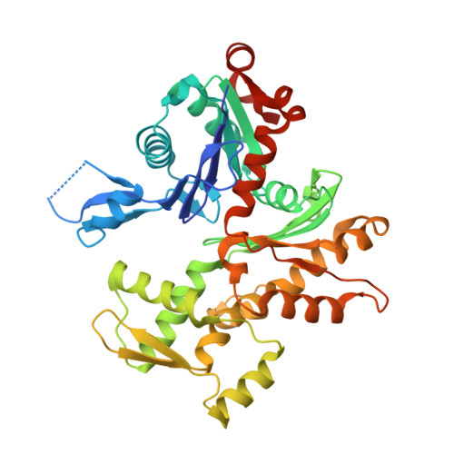
    6MGO, 6W7V

  • PubMed Abstract: 

    Cancer metastasis is a complex process involving highly motile tumor cells that breach tissue barriers, enter the bloodstream and lymphatic system, and disseminate throughout the body as circulating tumor cells. The primary cellular mechanism contributing to these critical events is the reorganization of the actin cytoskeleton. Mycalolide B (MycB) is an actin-targeting marine macrolide that can suppress proliferation, migration, and invasion of breast and ovarian cancer cells at low nanomolar doses. Through structure-activity relationship studies focused on the actin-binding tail region (C24-C35) of MycB, we identified a potent truncated derivative that inhibits polymerization of G-actin and severs F-actin by binding to actin's barbed end cleft. Biological analyses of this miniature MycB derivative demonstrate that it causes a rapid collapse of the actin cytoskeleton in ovarian cancer cells and impairs cancer cell motility and invasion of the extracellular matrix (ECM) by inhibiting invadopodia-mediated ECM degradation. These studies provide essential proof-of-principle for developing actin-targeting therapeutic agents to block cancer metastasis and establish a synthetically tractable barbed end-binding pharmacophore that can be further improved by adding targeting groups for precision drug design.


  • Organizational Affiliation
    • Department of Chemistry, Queen's University, 90 Bader Lane, Kingston ON K7L 3N6, Canada.

Macromolecule Content 

  • Total Structure Weight: 43.82 kDa 
  • Atom Count: 3,225 
  • Modeled Residue Count: 358 
  • Deposited Residue Count: 377 
  • Unique protein chains: 1

Macromolecules

Find similar proteins by:|  3D Structure
Entity ID: 1
MoleculeChains  Sequence LengthOrganismDetailsImage
Actin, alpha skeletal muscle377Oryctolagus cuniculusMutation(s): 0 
EC: 3.6.4
UniProt
Find proteins for P68135 (Oryctolagus cuniculus)
Explore P68135 
Go to UniProtKB:  P68135
Entity Groups
Sequence Clusters30% Identity50% Identity70% Identity90% Identity95% Identity100% Identity
UniProt GroupP68135
Sequence Annotations
Expand
Reference Sequence

Small Molecules

Ligands 5 Unique
IDChains Name / Formula / InChI Key2D Diagram3D Interactions
TFJ
(Subject of Investigation/LOI)

Query on TFJ


Download:Ideal Coordinates CCD File
F [auth A](1E,3R,4R,5S,6R,9S,10S,12S)-12-[(4-aminobutanoyl)oxy]-1-[ethyl(formyl)amino]-4,10-dimethoxy-3,5,9,13-tetramethyltetradec-1-en-6-yl (2R)-oxolane-2-carboxylate
C32 H58 N2 O8
DFAGFTKICVXUOP-RYYFKAAMSA-N
ATP

Query on ATP


Download:Ideal Coordinates CCD File
C [auth A]ADENOSINE-5'-TRIPHOSPHATE
C10 H16 N5 O13 P3
ZKHQWZAMYRWXGA-KQYNXXCUSA-N
LAB

Query on LAB


Download:Ideal Coordinates CCD File
D [auth A]LATRUNCULIN B
C20 H29 N O5 S
NSHPHXHGRHSMIK-JRIKCGFMSA-N
EDO

Query on EDO


Download:Ideal Coordinates CCD File
E [auth A],
G [auth A],
H [auth A]
1,2-ETHANEDIOL
C2 H6 O2
LYCAIKOWRPUZTN-UHFFFAOYSA-N
CA

Query on CA


Download:Ideal Coordinates CCD File
B [auth A]CALCIUM ION
Ca
BHPQYMZQTOCNFJ-UHFFFAOYSA-N

Experimental Data & Validation

Experimental Data

  • Method: X-RAY DIFFRACTION
  • Resolution: 1.70 Å
  • R-Value Free:  0.213 (Depositor), 0.210 (DCC) 
  • R-Value Work:  0.179 (Depositor), 0.174 (DCC) 
  • R-Value Observed: 0.181 (Depositor) 
Space Group: P 1 21 1
Unit Cell:
Length ( Å )Angle ( ˚ )
a = 37.763α = 90
b = 74.869β = 93.023
c = 61.197γ = 90
Software Package:
Software NamePurpose
PHENIXrefinement
PHENIXrefinement
PDB_EXTRACTdata extraction
XDSdata reduction
XDSdata scaling
PHASERphasing

Structure Validation

View Full Validation Report


Ligand Structure Quality Assessment 


Entry History 

& Funding Information

Deposition Data


Funding OrganizationLocationGrant Number
Canadian Institutes of Health Research (CIHR)Canada151976

Revision History  (Full details and data files)

  • Version 1.0: 2020-10-21
    Type: Initial release
  • Version 1.1: 2021-05-12
    Changes: Database references
  • Version 1.2: 2021-05-26
    Changes: Database references
  • Version 1.3: 2023-10-18
    Changes: Data collection, Database references, Refinement description