6W16

Crystal structure of a human metapneumovirus monomeric fusion protein complexed with 458 Fab


Experimental Data Snapshot

  • Method: X-RAY DIFFRACTION
  • Resolution: 3.10 Å
  • R-Value Free: 0.234 
  • R-Value Work: 0.190 
  • R-Value Observed: 0.193 

wwPDB Validation   3D Report Full Report


Ligand Structure Quality Assessment 


This is version 2.2 of the entry. See complete history


Literature

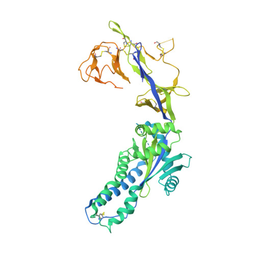
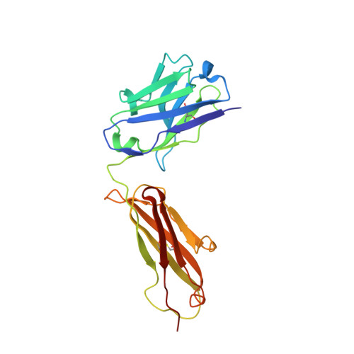
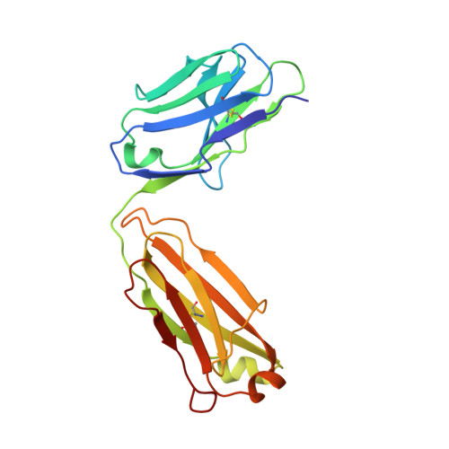
Antibody recognition of the Pneumovirus fusion protein trimer interface.

Huang, J.Diaz, D.Mousa, J.J.

(2020) PLoS Pathog 16: e1008942-e1008942

  • DOI: https://doi.org/10.1371/journal.ppat.1008942
  • Primary Citation of Related Structures:  
    6W16

  • PubMed Abstract: 

    Human metapneumovirus (hMPV) is a leading cause of viral respiratory infection in children, and can cause severe lower respiratory tract infection in infants, the elderly, and immunocompromised patients. However, there remain no licensed vaccines or specific treatments for hMPV infection. Although the hMPV fusion (F) protein is the sole target of neutralizing antibodies, the immunological properties of hMPV F remain poorly understood. To further define the humoral immune response to the hMPV F protein, we isolated two new human monoclonal antibodies (mAbs), MPV458 and MPV465. Both mAbs are neutralizing in vitro and were determined to target a unique antigenic site using competitive biolayer interferometry. We determined both MPV458 and MPV465 have higher affinity for monomeric hMPV F than trimeric hMPV F. MPV458 was co-crystallized with hMPV F, and the mAb primarily interacts with an alpha helix on the F2 region of the hMPV F protein. Surprisingly, the major epitope for MPV458 lies within the trimeric interface of the hMPV F protein, suggesting significant breathing of the hMPV F protein must occur for host immune recognition of the novel epitope. In addition, significant glycan interactions were observed with a somatically mutated light chain framework residue. The data presented identifies a novel epitope on the hMPV F protein for epitope-based vaccine design, and illustrates a new mechanism for human antibody neutralization of viral glycoproteins.


  • Organizational Affiliation

    Department of Infectious Diseases, College of Veterinary Medicine, University of Georgia, Athens, GA, United States of America.


Macromolecules
Find similar proteins by:  (by identity cutoff)  |  3D Structure
Entity ID: 1
MoleculeChains Sequence LengthOrganismDetailsImage
Fusion glycoprotein F0,Envelope glycoprotein fusionA [auth F]539human metapneumovirusHuman immunodeficiency virus 1Mutation(s): 0 
UniProt
Find proteins for M1E1E4 (Human immunodeficiency virus 1)
Explore M1E1E4 
Go to UniProtKB:  M1E1E4
Find proteins for Q6W8S4 (Human metapneumovirus)
Explore Q6W8S4 
Go to UniProtKB:  Q6W8S4
Entity Groups  
Sequence Clusters30% Identity50% Identity70% Identity90% Identity95% Identity100% Identity
UniProt GroupsM1E1E4Q6W8S4
Sequence Annotations
Expand
  • Reference Sequence
Find similar proteins by:  (by identity cutoff)  |  3D Structure
Entity ID: 2
MoleculeChains Sequence LengthOrganismDetailsImage
458 Fab heavy chainB [auth H]219Homo sapiensMutation(s): 0 
Entity Groups  
Sequence Clusters30% Identity50% Identity70% Identity90% Identity95% Identity100% Identity
Sequence Annotations
Expand
  • Reference Sequence
Find similar proteins by:  (by identity cutoff)  |  3D Structure
Entity ID: 3
MoleculeChains Sequence LengthOrganismDetailsImage
458 Fab light chainC [auth L]214Homo sapiensMutation(s): 0 
Entity Groups  
Sequence Clusters30% Identity50% Identity70% Identity90% Identity95% Identity100% Identity
Sequence Annotations
Expand
  • Reference Sequence
Oligosaccharides

Help

Entity ID: 4
MoleculeChains Length2D Diagram Glycosylation3D Interactions
alpha-D-mannopyranose-(1-3)-[alpha-D-mannopyranose-(1-6)]beta-D-mannopyranose-(1-4)-2-acetamido-2-deoxy-beta-D-glucopyranose-(1-4)-2-acetamido-2-deoxy-beta-D-glucopyranoseD [auth A]5N-Glycosylation
Glycosylation Resources
GlyTouCan:  G22768VO
GlyCosmos:  G22768VO
GlyGen:  G22768VO
Small Molecules
Ligands 1 Unique
IDChains Name / Formula / InChI Key2D Diagram3D Interactions
NAG
Query on NAG

Download Ideal Coordinates CCD File 
E [auth F]2-acetamido-2-deoxy-beta-D-glucopyranose
C8 H15 N O6
OVRNDRQMDRJTHS-FMDGEEDCSA-N
Experimental Data & Validation

Experimental Data

  • Method: X-RAY DIFFRACTION
  • Resolution: 3.10 Å
  • R-Value Free: 0.234 
  • R-Value Work: 0.190 
  • R-Value Observed: 0.193 
  • Space Group: P 65
Unit Cell:
Length ( Å )Angle ( ˚ )
a = 128.489α = 90
b = 128.489β = 90
c = 188.352γ = 120
Software Package:
Software NamePurpose
PHENIXrefinement
XDSdata reduction
Aimlessdata scaling
PHASERphasing

Structure Validation

View Full Validation Report


Ligand Structure Quality Assessment 


Entry History & Funding Information

Deposition Data


Funding OrganizationLocationGrant Number
National Institutes of Health/National Institute Of Allergy and Infectious Diseases (NIH/NIAID)United StatesR01AI14386

Revision History  (Full details and data files)

  • Version 1.0: 2020-06-03
    Type: Initial release
  • Version 2.0: 2020-07-29
    Type: Remediation
    Reason: Carbohydrate remediation
    Changes: Atomic model, Data collection, Derived calculations, Structure summary
  • Version 2.1: 2021-02-10
    Changes: Database references, Structure summary
  • Version 2.2: 2023-10-18
    Changes: Data collection, Database references, Refinement description