6VX6 | pdb_00006vx6

bestrophin-2 Ca2+-bound state (250 nM Ca2+)


Experimental Data Snapshot

  • Method: ELECTRON MICROSCOPY
  • Resolution: 3.00 Å
  • Aggregation State: PARTICLE 
  • Reconstruction Method: SINGLE PARTICLE 

wwPDB Validation 3D Report Full Report

Validation slider image for 6VX6

This is version 1.2 of the entry. See complete history

Literature

Structural and functional characterization of the bestrophin-2 anion channel.

Owji, A.P.Zhao, Q.Ji, C.Kittredge, A.Hopiavuori, A.Fu, Z.Ward, N.Clarke, O.B.Shen, Y.Zhang, Y.Hendrickson, W.A.Yang, T.

(2020) Nat Struct Mol Biol 27: 382-391

  • DOI: https://doi.org/10.1038/s41594-020-0402-z
  • Primary Citation Related Structures: 
    6VX5, 6VX6, 6VX7, 6VX8, 6VX9

  • PubMed Abstract: 

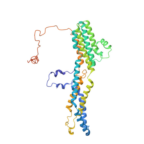
    The bestrophin family of calcium (Ca 2+ )-activated chloride (Cl - ) channels, which mediate the influx and efflux of monovalent anions in response to the levels of intracellular Ca 2+ , comprises four members in mammals (bestrophin 1-4). Here we report cryo-EM structures of bovine bestrophin-2 (bBest2) bound and unbound by Ca 2+ at 2.4- and 2.2-Å resolution, respectively. The bBest2 structure highlights four previously underappreciated pore-lining residues specifically conserved in Best2 but not in Best1, illustrating the differences between these paralogs. Structure-inspired electrophysiological analysis reveals that, although the channel is sensitive to Ca 2+ , it has substantial Ca 2+ -independent activity for Cl - , reflecting the opening at the cytoplasmic restriction of the ion conducting pathway even when Ca 2+ is absent. Moreover, the ion selectivity of bBest2 is controlled by multiple residues, including those involved in gating.


  • Organizational Affiliation
    • Department of Pharmacology, Columbia University, New York, NY, USA.

Macromolecule Content 

  • Total Structure Weight: 237.36 kDa 
  • Atom Count: 15,061 
  • Modeled Residue Count: 1,830 
  • Deposited Residue Count: 2,050 
  • Unique protein chains: 1

Macromolecules

Find similar proteins by:|  3D Structure
Entity ID: 1
MoleculeChains  Sequence LengthOrganismDetailsImage
BestrophinA [auth E],
B [auth A],
C [auth B],
D [auth C],
E [auth D]
410Bos taurusMutation(s): 0 
Gene Names: BEST2
Membrane Entity: Yes 
UniProt
Find proteins for E1BF86 (Bos taurus)
Explore E1BF86 
Go to UniProtKB:  E1BF86
Entity Groups
Sequence Clusters30% Identity50% Identity70% Identity90% Identity95% Identity100% Identity
UniProt GroupE1BF86
Sequence Annotations
Expand
Reference Sequence

Experimental Data & Validation

Experimental Data

  • Method: ELECTRON MICROSCOPY
  • Resolution: 3.00 Å
  • Aggregation State: PARTICLE 
  • Reconstruction Method: SINGLE PARTICLE 
EM Software:
TaskSoftware PackageVersion
RECONSTRUCTIONcryoSPARC

Structure Validation

View Full Validation Report


Entry History 

& Funding Information

Deposition Data


Funding OrganizationLocationGrant Number
National Institutes of Health/National Eye Institute (NIH/NEI)United States1F31EY030763
National Institutes of Health/National Eye Institute (NIH/NEI)United StatesEY025290
National Institutes of Health/National Institute of General Medical Sciences (NIH/NIGMS)United States7R01GM127652
National Institutes of Health/National Institute of General Medical Sciences (NIH/NIGMS)United States3R01GM107462

Revision History  (Full details and data files)

  • Version 1.0: 2020-04-08
    Type: Initial release
  • Version 1.1: 2020-04-22
    Changes: Database references
  • Version 1.2: 2024-03-06
    Changes: Data collection, Database references, Derived calculations