6VUA | pdb_00006vua

X-ray structure of human CD38 catalytic domain with 2'-Cl-araNAD+


Experimental Data Snapshot

  • Method: X-RAY DIFFRACTION
  • Resolution: 1.50 Å
  • R-Value Free: 
    0.190 (Depositor), 0.194 (DCC) 
  • R-Value Work: 
    0.166 (Depositor), 0.172 (DCC) 
  • R-Value Observed: 
    0.168 (Depositor) 

Starting Model: experimental
View more details

wwPDB Validation 3D Report Full Report

Validation slider image for 6VUA

Ligand Structure Quality Assessment 


This is version 1.3 of the entry. See complete history

Literature

Synthesis of site-specific antibody-drug conjugates by ADP-ribosyl cyclases.

Dai, Z.Zhang, X.N.Nasertorabi, F.Cheng, Q.Li, J.Katz, B.B.Smbatyan, G.Pei, H.Louie, S.G.Lenz, H.J.Stevens, R.C.Zhang, Y.

(2020) Sci Adv 6: eaba6752-eaba6752

  • DOI: https://doi.org/10.1126/sciadv.aba6752
  • Primary Citation Related Structures: 
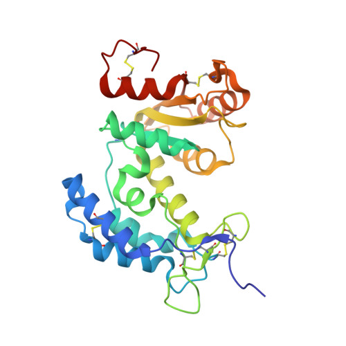
    6VUA

  • PubMed Abstract: 

    Most of the current antibody-drug conjugates (ADCs) in clinic are heterogeneous mixtures. To produce homogeneous ADCs, established procedures often require multiple steps or long reaction times. The introduced mutations or foreign sequences may cause high immunogenicity. Here, we explore a new concept of transforming CD38 enzymatic activity into a facile approach for generating site-specific ADCs. This was achieved through coupling bifunctional antibody-CD38 fusion proteins with designer dinucleotide-based covalent inhibitors with stably attached payloads. The resulting adenosine diphosphate-ribosyl cyclase-enabled ADC (ARC-ADC) with a drug-to-antibody ratio of 2 could be rapidly generated through single-step conjugation. The generated ARC-ADC targeting human epidermal growth factor receptor 2 (HER2) displays excellent stability and potency against HER2-positive breast cancer both in vitro and in vivo. This proof-of-concept study demonstrates a new strategy for production of site-specific ADCs. It may provide a general approach for the development of a novel class of ADCs with potentially enhanced properties.


  • Organizational Affiliation
    • Department of Pharmacology and Pharmaceutical Sciences, School of Pharmacy, University of Southern California, Los Angeles, CA 90089, USA.

Macromolecule Content 

  • Total Structure Weight: 61.95 kDa 
  • Atom Count: 4,973 
  • Modeled Residue Count: 506 
  • Deposited Residue Count: 514 
  • Unique protein chains: 1

Macromolecules

Find similar proteins by:|  3D Structure
Entity ID: 1
MoleculeChains  Sequence LengthOrganismDetailsImage
ADP-ribosyl cyclase/cyclic ADP-ribose hydrolase 1
A, B
257Homo sapiensMutation(s): 4 
Gene Names: CD38
EC: 3.2.2.6 (PDB Primary Data), 2.4.99.20 (PDB Primary Data), 3.2.2 (UniProt)
UniProt & NIH Common Fund Data Resources
Find proteins for P28907 (Homo sapiens)
Explore P28907 
Go to UniProtKB:  P28907
PHAROS:  P28907
GTEx:  ENSG00000004468 
Entity Groups
Sequence Clusters30% Identity50% Identity70% Identity90% Identity95% Identity100% Identity
UniProt GroupP28907
Sequence Annotations
Expand
Reference Sequence

Small Molecules

Ligands 6 Unique
IDChains Name / Formula / InChI Key2D Diagram3D Interactions
ROJ
(Subject of Investigation/LOI)

Query on ROJ


Download:Ideal Coordinates CCD File
C [auth A],
J [auth B]
[[(2~{R},3~{S},4~{R},5~{R})-5-(6-aminopurin-9-yl)-3,4-bis(oxidanyl)oxolan-2-yl]methoxy-oxidanyl-phosphoryl] [(2~{R},3~{R},4~{R})-4-chloranyl-3-oxidanyl-oxolan-2-yl]methyl hydrogen phosphate
C15 H22 Cl N5 O12 P2
JYKQNUHEQVWWJW-WFCIOUKESA-N
PGE

Query on PGE


Download:Ideal Coordinates CCD File
H [auth A]TRIETHYLENE GLYCOL
C6 H14 O4
ZIBGPFATKBEMQZ-UHFFFAOYSA-N
MPD

Query on MPD


Download:Ideal Coordinates CCD File
G [auth A],
M [auth B],
N [auth B]
(4S)-2-METHYL-2,4-PENTANEDIOL
C6 H14 O2
SVTBMSDMJJWYQN-YFKPBYRVSA-N
PEG

Query on PEG


Download:Ideal Coordinates CCD File
F [auth A],
I [auth A],
O [auth B],
P [auth B],
Q [auth B]
DI(HYDROXYETHYL)ETHER
C4 H10 O3
MTHSVFCYNBDYFN-UHFFFAOYSA-N
PO4

Query on PO4


Download:Ideal Coordinates CCD File
E [auth A],
L [auth B]
PHOSPHATE ION
O4 P
NBIIXXVUZAFLBC-UHFFFAOYSA-K
K

Query on K


Download:Ideal Coordinates CCD File
D [auth A],
K [auth B]
POTASSIUM ION
K
NPYPAHLBTDXSSS-UHFFFAOYSA-N

Experimental Data & Validation

Experimental Data

  • Method: X-RAY DIFFRACTION
  • Resolution: 1.50 Å
  • R-Value Free:  0.190 (Depositor), 0.194 (DCC) 
  • R-Value Work:  0.166 (Depositor), 0.172 (DCC) 
  • R-Value Observed: 0.168 (Depositor) 
Space Group: P 41 2 2
Unit Cell:
Length ( Å )Angle ( ˚ )
a = 114.762α = 90
b = 114.762β = 90
c = 97.154γ = 90
Software Package:
Software NamePurpose
XDSdata reduction
SCALAdata scaling
PHASERphasing
REFMACrefinement
PDB_EXTRACTdata extraction

Structure Validation

View Full Validation Report


Ligand Structure Quality Assessment 


Entry History 

& Funding Information

Deposition Data


Funding OrganizationLocationGrant Number
Other privateUnited StatesUniversity of Southern California Start-Up Fund for New Faculty and Sharon L. Cockrell Cancer Research Fund
Other privateUnited StatesThe V Foundation for Cancer Research V2016-021
Other privateUnited StatesSTOP CANCER Research Career Development Award
Other privateUnited StatesPhRMA Foundation Research Starter Grant in Translational Medicine and Therapeutics

Revision History  (Full details and data files)

  • Version 1.0: 2020-06-03
    Type: Initial release
  • Version 1.1: 2020-06-24
    Changes: Database references
  • Version 1.2: 2023-10-11
    Changes: Data collection, Database references, Derived calculations, Refinement description
  • Version 1.3: 2024-10-23
    Changes: Structure summary