6VPG

TPX2 residues 7-20 fused to Aurora A residues 116-389 in complex with AMP-PNP

  • Classification: TRANSFERASE
  • Organism(s): Homo sapiens
  • Expression System: Escherichia coli
  • Mutation(s): No 

  • Deposited: 2020-02-03 Released: 2020-08-05 
  • Deposition Author(s): Lim, D.C., Yaffe, M.B.
  • Funding Organization(s): National Institutes of Health/National Institute of Environmental Health Sciences (NIH/NIEHS), National Institutes of Health/National Institute of General Medical Sciences (NIH/NIGMS)

Experimental Data Snapshot

  • Method: X-RAY DIFFRACTION
  • Resolution: 2.64 Å
  • R-Value Free: 0.245 
  • R-Value Work: 0.199 
  • R-Value Observed: 0.204 

wwPDB Validation   3D Report Full Report


Ligand Structure Quality Assessment 


This is version 1.1 of the entry. See complete history


Literature

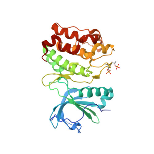
Redox priming promotes Aurora A activation during mitosis.

Lim, D.C.Joukov, V.Rettenmaier, T.J.Kumagai, A.Dunphy, W.G.Wells, J.A.Yaffe, M.B.

(2020) Sci Signal 13

  • DOI: https://doi.org/10.1126/scisignal.abb6707
  • Primary Citation of Related Structures:  
    6VPG, 6VPH, 6VPI, 6VPJ, 6VPL, 6VPM, 6XKA

  • PubMed Abstract: 

    Cell cycle-dependent redox changes can mediate transient covalent modifications of cysteine thiols to modulate the activities of regulatory kinases and phosphatases. Our previously reported finding that protein cysteine oxidation is increased during mitosis relative to other cell cycle phases suggests that redox modifications could play prominent roles in regulating mitotic processes. The Aurora family of kinases and their downstream targets are key components of the cellular machinery that ensures the proper execution of mitosis and the accurate segregation of chromosomes to daughter cells. In this study, x-ray crystal structures of the Aurora A kinase domain delineate redox-sensitive cysteine residues that, upon covalent modification, can allosterically regulate kinase activity and oligomerization state. We showed in both Xenopus laevis egg extracts and mammalian cells that a conserved cysteine residue within the Aurora A activation loop is crucial for Aurora A activation by autophosphorylation. We further showed that covalent disulfide adducts of this residue promote autophosphorylation of the Aurora A kinase domain. These findings reveal a potential mechanistic link between Aurora A activation and changes in the intracellular redox state during mitosis and provide insights into how novel small-molecule inhibitors may be developed to target specific subpopulations of Aurora A.


  • Organizational Affiliation

    MIT Center for Precision Cancer Medicine, Koch Institute for Integrative Cancer Research, and Departments of Biological Engineering and Biology, Massachusetts Institute of Technology, Cambridge, MA 02139, USA. danlim@mit.edu myaffe@mit.edu.


Macromolecules
Find similar proteins by:  (by identity cutoff)  |  3D Structure
Entity ID: 1
MoleculeChains Sequence LengthOrganismDetailsImage
TPX2 fragment - Aurora A kinase domain fusion292Homo sapiensMutation(s): 0 
Gene Names: 
TPX2C20orf1C20orf2DIL2HCA519AURKAAIKAIRK1ARK1AURAAYK1BTAKIAK1STK15STK6

EC: 2.7.11.1
UniProt & NIH Common Fund Data Resources
Find proteins for Q9ULW0 (Homo sapiens)
Explore Q9ULW0 
Go to UniProtKB:  Q9ULW0
PHAROS:  Q9ULW0
GTEx:  ENSG00000088325 
Find proteins for O14965 (Homo sapiens)
Explore O14965 
Go to UniProtKB:  O14965
PHAROS:  O14965
GTEx:  ENSG00000087586 
Entity Groups  
Sequence Clusters30% Identity50% Identity70% Identity90% Identity95% Identity100% Identity
UniProt GroupsO14965Q9ULW0
Sequence Annotations
Expand
  • Reference Sequence
Small Molecules
Experimental Data & Validation

Experimental Data

  • Method: X-RAY DIFFRACTION
  • Resolution: 2.64 Å
  • R-Value Free: 0.245 
  • R-Value Work: 0.199 
  • R-Value Observed: 0.204 
  • Space Group: P 41 21 2
Unit Cell:
Length ( Å )Angle ( ˚ )
a = 83.562α = 90
b = 83.562β = 90
c = 114.516γ = 90
Software Package:
Software NamePurpose
PHENIXrefinement
SCALEPACKdata scaling
AMoREphasing
PDB_EXTRACTdata extraction
DENZOdata reduction

Structure Validation

View Full Validation Report


Ligand Structure Quality Assessment 


Entry History & Funding Information

Deposition Data


Funding OrganizationLocationGrant Number
National Institutes of Health/National Institute of Environmental Health Sciences (NIH/NIEHS)United StatesES015339
National Institutes of Health/National Institute of Environmental Health Sciences (NIH/NIEHS)United StatesES028374
National Institutes of Health/National Institute of Environmental Health Sciences (NIH/NIEHS)United StatesES020466
National Institutes of Health/National Institute of General Medical Sciences (NIH/NIGMS)United StatesGM104047

Revision History  (Full details and data files)

  • Version 1.0: 2020-08-05
    Type: Initial release
  • Version 1.1: 2023-10-11
    Changes: Data collection, Database references, Refinement description