6VP6

Cryo-EM structure of the C-terminal half of the Parkinson's Disease-linked protein Leucine Rich Repeat Kinase 2 (LRRK2)


Experimental Data Snapshot

  • Method: ELECTRON MICROSCOPY
  • Resolution: 3.47 Å
  • Aggregation State: PARTICLE 
  • Reconstruction Method: SINGLE PARTICLE 

wwPDB Validation   3D Report Full Report


This is version 1.2 of the entry. See complete history


Literature

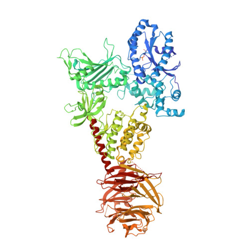
Structure of LRRK2 in Parkinson's disease and model for microtubule interaction.

Deniston, C.K.Salogiannis, J.Mathea, S.Snead, D.M.Lahiri, I.Matyszewski, M.Donosa, O.Watanabe, R.Bohning, J.Shiau, A.K.Knapp, S.Villa, E.Reck-Peterson, S.L.Leschziner, A.E.

(2020) Nature 588: 344-349

  • DOI: https://doi.org/10.1038/s41586-020-2673-2
  • Primary Citation of Related Structures:  
    6VNO, 6VP6, 6VP7, 6VP8

  • PubMed Abstract: 

    Leucine-rich repeat kinase 2 (LRRK2) is the most commonly mutated gene in familial Parkinson's disease 1 and is also linked to its idiopathic form 2 . LRRK2 has been proposed to function in membrane trafficking 3 and colocalizes with microtubules 4 . Despite the fundamental importance of LRRK2 for understanding and treating Parkinson's disease, structural information on the enzyme is limited. Here we report the structure of the catalytic half of LRRK2, and an atomic model of microtubule-associated LRRK2 built using a reported cryo-electron tomography in situ structure 5 . We propose that the conformation of the LRRK2 kinase domain regulates its interactions with microtubules, with a closed conformation favouring oligomerization on microtubules. We show that the catalytic half of LRRK2 is sufficient for filament formation and blocks the motility of the microtubule-based motors kinesin 1 and cytoplasmic dynein 1 in vitro. Kinase inhibitors that stabilize an open conformation relieve this interference and reduce the formation of LRRK2 filaments in cells, whereas inhibitors that stabilize a closed conformation do not. Our findings suggest that LRRK2 can act as a roadblock for microtubule-based motors and have implications for the design of therapeutic LRRK2 kinase inhibitors.


  • Organizational Affiliation

    Department of Cellular and Molecular Medicine, University of California San Diego, La Jolla, CA, USA.


Macromolecules
Find similar proteins by:  (by identity cutoff)  |  3D Structure
Entity ID: 1
MoleculeChains Sequence LengthOrganismDetailsImage
Leucine-rich repeat serine/threonine-protein kinase 2
A, B, C
1,201Homo sapiensMutation(s): 0 
Gene Names: LRRK2PARK8
EC: 2.7.11.1 (PDB Primary Data), 3.6.5 (PDB Primary Data)
UniProt & NIH Common Fund Data Resources
Find proteins for Q5S007 (Homo sapiens)
Explore Q5S007 
Go to UniProtKB:  Q5S007
PHAROS:  Q5S007
GTEx:  ENSG00000188906 
Entity Groups  
Sequence Clusters30% Identity50% Identity70% Identity90% Identity95% Identity100% Identity
UniProt GroupQ5S007
Sequence Annotations
Expand
  • Reference Sequence
Small Molecules
Ligands 2 Unique
IDChains Name / Formula / InChI Key2D Diagram3D Interactions
GDP
Query on GDP

Download Ideal Coordinates CCD File 
D [auth A]GUANOSINE-5'-DIPHOSPHATE
C10 H15 N5 O11 P2
QGWNDRXFNXRZMB-UUOKFMHZSA-N
MG
Query on MG

Download Ideal Coordinates CCD File 
E [auth A]MAGNESIUM ION
Mg
JLVVSXFLKOJNIY-UHFFFAOYSA-N
Modified Residues  1 Unique
IDChains TypeFormula2D DiagramParent
TPO
Query on TPO
A, B, C
L-PEPTIDE LINKINGC4 H10 N O6 PTHR
Experimental Data & Validation

Experimental Data

  • Method: ELECTRON MICROSCOPY
  • Resolution: 3.47 Å
  • Aggregation State: PARTICLE 
  • Reconstruction Method: SINGLE PARTICLE 
EM Software:
TaskSoftware PackageVersion
MODEL REFINEMENTRosetta3
RECONSTRUCTIONRELION3
RECONSTRUCTIONcryoSPARC2

Structure Validation

View Full Validation Report


Entry History & Funding Information

Deposition Data


Funding OrganizationLocationGrant Number
Michael J. Fox FoundationUnited States11425
Michael J. Fox FoundationUnited States11425.02
National Institutes of Health/National Institute of General Medical Sciences (NIH/NIGMS)United StatesR01GM107214

Revision History  (Full details and data files)

  • Version 1.0: 2020-08-26
    Type: Initial release
  • Version 1.1: 2020-09-02
    Changes: Database references
  • Version 1.2: 2020-12-23
    Changes: Database references