6VOY

Cryo-EM structure of HTLV-1 instasome


Experimental Data Snapshot

  • Method: ELECTRON MICROSCOPY
  • Resolution: 3.70 Å
  • Aggregation State: PARTICLE 
  • Reconstruction Method: SINGLE PARTICLE 

wwPDB Validation   3D Report Full Report


This is version 1.0 of the entry. See complete history


Literature

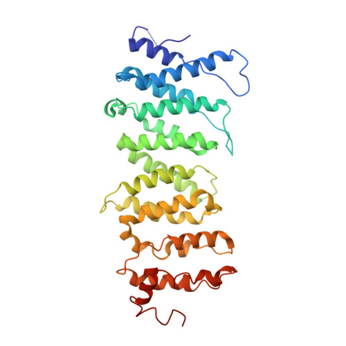
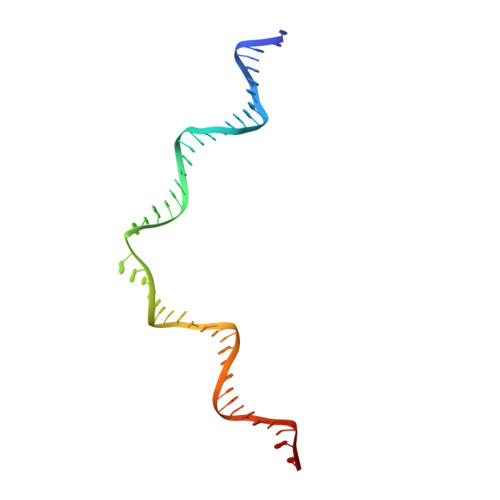
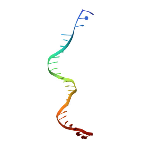
Structural basis of host protein hijacking in human T-cell leukemia virus integration.

Bhatt, V.Shi, K.Salamango, D.J.Moeller, N.H.Pandey, K.K.Bera, S.Bohl, T.E.Kurniawan, F.Orellana, K.Zhang, W.Grandgenett, D.P.Harris, R.S.Sundborger-Lunna, A.C.Aihara, H.

(2020) Nat Commun 11: 3121-3121

  • DOI: https://doi.org/10.1038/s41467-020-16963-6
  • Primary Citation of Related Structures:  
    6VOY

  • PubMed Abstract: 
  • Integration of the reverse-transcribed viral DNA into host chromosomes is a critical step in the life-cycle of retroviruses, including an oncogenic delta(δ)-retrovirus human T-cell leukemia virus type-1 (HTLV-1). Retroviral integrase forms a higher order nucleoprotein assembly (intasome) to catalyze the integration reaction, in which the roles of host factors remain poorly understood ...

    Integration of the reverse-transcribed viral DNA into host chromosomes is a critical step in the life-cycle of retroviruses, including an oncogenic delta(δ)-retrovirus human T-cell leukemia virus type-1 (HTLV-1). Retroviral integrase forms a higher order nucleoprotein assembly (intasome) to catalyze the integration reaction, in which the roles of host factors remain poorly understood. Here, we use cryo-electron microscopy to visualize the HTLV-1 intasome at 3.7-Å resolution. The structure together with functional analyses reveal that the B56γ (B'γ) subunit of an essential host enzyme, protein phosphatase 2 A (PP2A), is repurposed as an integral component of the intasome to mediate HTLV-1 integration. Our studies reveal a key host-virus interaction underlying the replication of an important human pathogen and highlight divergent integration strategies of retroviruses.


    Organizational Affiliation

    Institute for Molecular Virology, University of Minnesota, 515 Delaware Street S.E., Minneapolis, MN, 55455, USA. aihar001@umn.edu.


Macromolecules

Find similar proteins by:  (by identity cutoff)  |  3D Structure
Entity ID: 1
MoleculeChainsSequence LengthOrganismDetailsImage
DNA-binding protein 7d
A, B, C, D
390Saccharolobus solfataricus P2Human T-cell leukemia virus type I
This entity is chimeric
Mutation(s): 1 
Gene Names: sso7dsso7d-1SSO10610pol
EC: 3.4.23 (UniProt), 2.7.7.49 (UniProt), 2.7.7.7 (UniProt), 3.1.26.4 (UniProt), 2.7.7 (UniProt), 3.1 (UniProt)
UniProt
Find proteins for P39476 (Saccharolobus solfataricus (strain ATCC 35092 / DSM 1617 / JCM 11322 / P2))
Explore P39476 
Go to UniProtKB:  P39476
Find proteins for P14078 (Human T-cell leukemia virus 1 (isolate Caribbea HS-35 subtype A))
Explore P14078 
Go to UniProtKB:  P14078
Entity Groups  
Sequence Clusters30% Identity50% Identity70% Identity90% Identity95% Identity100% Identity
UniProt GroupsP14078P39476
Protein Feature View
Expand
  • Reference Sequence
Find similar proteins by:  (by identity cutoff)  |  3D Structure
Entity ID: 2
MoleculeChainsSequence LengthOrganismDetailsImage
Serine/threonine-protein phosphatase 2A 56 kDa regulatory subunit gamma isoform
E, F
343Homo sapiensMutation(s): 0 
Gene Names: PPP2R5CKIAA0044
UniProt & NIH Common Fund Data Resources
Find proteins for Q13362 (Homo sapiens)
Explore Q13362 
Go to UniProtKB:  Q13362
PHAROS:  Q13362
Entity Groups  
Sequence Clusters30% Identity50% Identity70% Identity90% Identity95% Identity100% Identity
UniProt GroupQ13362
Protein Feature View
Expand
  • Reference Sequence
Find similar nucleic acids by:  (by identity cutoff)  |  3D Structure
Entity ID: 3
MoleculeChainsLengthOrganismImage
DNA (5'-D(P*AP*CP*AP*CP*AP*CP*TP*TP*GP*AP*CP*TP*AP*GP*GP*GP*TP*G)-3')G [auth I],
J [auth L]
49Homo sapiens
Protein Feature View
Expand
  • Reference Sequence

Find similar nucleic acids by:  Sequence   |   3D Structure  

Entity ID: 4
MoleculeChainsLengthOrganismImage
DNA (25-MER)H [auth J],
K [auth M]
25Human T-cell leukemia virus type I
Protein Feature View
Expand
  • Reference Sequence

Find similar nucleic acids by:  Sequence   |   3D Structure  

Entity ID: 5
MoleculeChainsLengthOrganismImage
DNA (5'-D(P*AP*CP*AP*CP*AP*CP*TP*TP*GP*AP*CP*TP*AP*GP*GP*GP*TP*G)-3')I [auth K],
L [auth N]
20Human T-cell leukemia virus type I
Protein Feature View
Expand
  • Reference Sequence
Experimental Data & Validation

Experimental Data

  • Method: ELECTRON MICROSCOPY
  • Resolution: 3.70 Å
  • Aggregation State: PARTICLE 
  • Reconstruction Method: SINGLE PARTICLE 
EM Software:
TaskSoftware PackageVersion
RECONSTRUCTIONRELION3.08

Structure Validation

View Full Validation Report


Entry History & Funding Information

Deposition Data


Funding OrganizationLocationGrant Number
National Institutes of Health/National Institute of General Medical Sciences (NIH/NIGMS)United States--

Revision History  (Full details and data files)

  • Version 1.0: 2020-07-01
    Type: Initial release