


Crystal structure of DPO4 extension past 8-oxoadenine (oxoA) and dT
Jung, H., Lee, S.To be published.
Experimental Data Snapshot
Starting Model: experimental
View more details
Entity ID: 3 | |||||
|---|---|---|---|---|---|
| Molecule | Chains | Sequence Length | Organism | Details | Image |
| DNA polymerase IV | C [auth A], F [auth D], I [auth G], L [auth J] | 341 | Saccharolobus solfataricus | Mutation(s): 0 Gene Names: dbh, SSOP1_2570, SULA_0242, SULB_0243, SULC_0242, SULG_01230, SULH_01230, SULI_01230, SULM_01230, SULN_01230... EC: 2.7.7.7 | 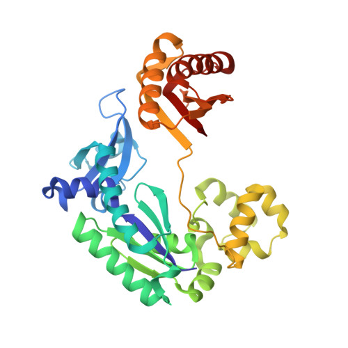 |
UniProt | |||||
Entity Groups | |||||
| Sequence Clusters | 30% Identity50% Identity70% Identity90% Identity95% Identity100% Identity | ||||
| UniProt Group | Q97W02 | ||||
Sequence AnnotationsExpand | |||||
Reference Sequence | |||||
Entity ID: 1 | ||||
| Molecule | Chains | Length | Organism | Image |
|---|---|---|---|---|
| DNA (5'-D(P*GP*GP*GP*GP*GP*AP*AP*GP*GP*AP*TP*TP*CP*T)-3') | A [auth P], D [auth B], G [auth E], J [auth H] | 14 | Homo sapiens | 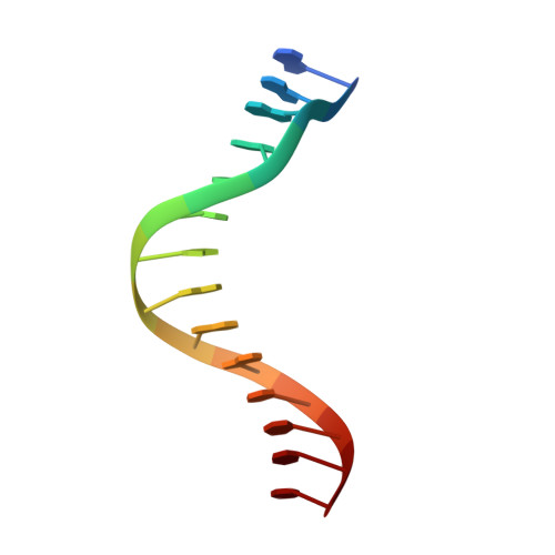 |
Sequence AnnotationsExpand | ||||
Reference Sequence | ||||
Entity ID: 2 | ||||
| Molecule | Chains | Length | Organism | Image |
|---|---|---|---|---|
| DNA (5'-D(*TP*TP*CP*AP*T*(R5M)P*GP*AP*AP*TP*CP*CP*TP*TP*CP*CP*CP*CP*C)-3') | B [auth T], E [auth C], H [auth F], K [auth I] | 19 | Homo sapiens | 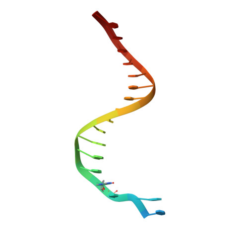 |
Sequence AnnotationsExpand | ||||
Reference Sequence | ||||
| Ligands 2 Unique | |||||
|---|---|---|---|---|---|
| ID | Chains | Name / Formula / InChI Key | 2D Diagram | 3D Interactions | |
| DZ4 (Subject of Investigation/LOI) Download:Ideal Coordinates CCD File | M [auth A], P [auth D], S [auth G], V [auth J] | 2'-deoxy-5'-O-[(R)-hydroxy{[(R)-hydroxy(phosphonooxy)phosphoryl]amino}phosphoryl]adenosine C10 H17 N6 O11 P3 WKIPJDSLGCBQCU-RRKCRQDMSA-N |  | ||
| MG Download:Ideal Coordinates CCD File | N [auth A] O [auth A] Q [auth D] R [auth D] T [auth G] | MAGNESIUM ION Mg JLVVSXFLKOJNIY-UHFFFAOYSA-N |  | ||
| Length ( Å ) | Angle ( ˚ ) |
|---|---|
| a = 53.255 | α = 90.03 |
| b = 98.984 | β = 89.99 |
| c = 102.875 | γ = 89.92 |
| Software Name | Purpose |
|---|---|
| PHENIX | refinement |
| HKL-2000 | data reduction |
| HKL-2000 | data scaling |
| MOLREP | phasing |


