6VMZ | pdb_00006vmz

Crystal Structure of a H5N1 influenza virus hemagglutinin with CBS1117


Experimental Data Snapshot

  • Method: X-RAY DIFFRACTION
  • Resolution: 2.20 Å
  • R-Value Free: 
    0.252 (Depositor), 0.251 (DCC) 
  • R-Value Work: 
    0.208 (Depositor), 0.211 (DCC) 
  • R-Value Observed: 
    0.210 (Depositor) 

Starting Model: experimental
View more details

wwPDB Validation 3D Report Full Report

Validation slider image for 6VMZ

Ligand Structure Quality Assessment 


This is version 1.4 of the entry. See complete history

Literature

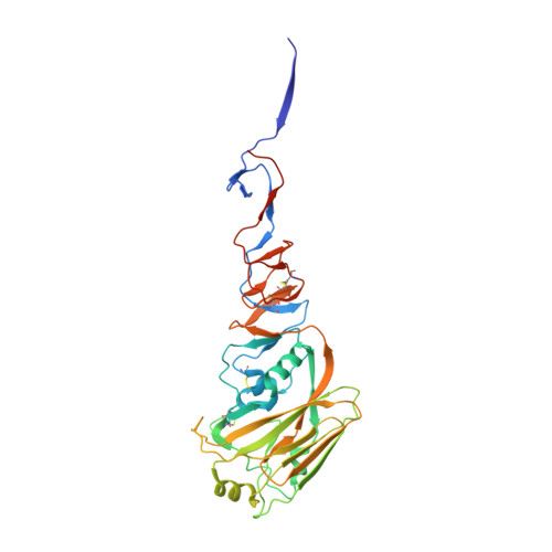
Structure of avian influenza hemagglutinin in complex with a small molecule entry inhibitor.

Antanasijevic, A.Durst, M.A.Cheng, H.Gaisina, I.N.Perez, J.T.Manicassamy, B.Rong, L.Lavie, A.Caffrey, M.

(2020) Life Sci Alliance 3

  • DOI: https://doi.org/10.26508/lsa.202000724
  • Primary Citation Related Structures: 
    6VMZ

  • PubMed Abstract: 

    HA plays a critical role in influenza infection and, thus HA is a potential target for antivirals. Recently, our laboratories have described a novel fusion inhibitor, termed CBS1117, with EC 50 ∼3 μM against group 1 HA. In this work, we characterize the binding properties of CBS1117 to avian H5 HA by x-ray crystallography, NMR, and mutagenesis. The x-ray structure of the complex shows that the compound binds near the HA fusion peptide, a region that plays a critical role in HA-mediated fusion. NMR studies demonstrate binding of CBS1117 to H5 HA in solution and show extensive hydrophobic contacts between the compound and HA surface. Mutagenesis studies further support the location of the compound binding site proximal to the HA fusion peptide and identify additional amino acids that are important to compound binding. Together, this work gives new insights into the CBS1117 mechanism of action and can be exploited to further optimize this compound and better understand the group specific activity of small-molecule inhibitors of HA-mediated entry.


  • Organizational Affiliation
    • Department of Biochemistry and Molecular Genetics, University of Illinois at Chicago, Chicago, IL, USA.

Macromolecule Content 

  • Total Structure Weight: 179.2 kDa 
  • Atom Count: 12,247 
  • Modeled Residue Count: 1,454 
  • Deposited Residue Count: 1,545 
  • Unique protein chains: 2

Macromolecules

Find similar proteins by:|  3D Structure
Entity ID: 1
MoleculeChains  Sequence LengthOrganismDetailsImage
HemagglutininA,
B [auth C],
C [auth E]
334Influenza A virus (A/chicken/Vietnam/4/2003(H5N1))Mutation(s): 0 
Gene Names: HA
UniProt
Find proteins for Q1KHJ8 (Influenza A virus)
Explore Q1KHJ8 
Go to UniProtKB:  Q1KHJ8
Entity Groups
Sequence Clusters30% Identity50% Identity70% Identity90% Identity95% Identity100% Identity
UniProt GroupQ1KHJ8
Glycosylation
Glycosylation Sites: 2
Sequence Annotations
Expand
Reference Sequence
Find similar proteins by:|  3D Structure
Entity ID: 2
MoleculeChains  Sequence LengthOrganismDetailsImage
HemagglutininD [auth B],
E [auth D],
F
181Influenza A virus (A/chicken/Vietnam/30/2003(H5N1))Mutation(s): 0 
Gene Names: HA
UniProt
Find proteins for Q1KHK7 (Influenza A virus)
Explore Q1KHK7 
Go to UniProtKB:  Q1KHK7
Entity Groups
Sequence Clusters30% Identity50% Identity70% Identity90% Identity95% Identity100% Identity
UniProt GroupQ1KHK7
Sequence Annotations
Expand
Reference Sequence

Small Molecules

Ligands 2 Unique
IDChains Name / Formula / InChI Key2D Diagram3D Interactions
R3P
(Subject of Investigation/LOI)

Query on R3P


Download:Ideal Coordinates CCD File
J [auth A],
N [auth C],
Q [auth E]
2,6-dichloro-N-[1-(propan-2-yl)piperidin-4-yl]benzamide
C15 H20 Cl2 N2 O
OUZZSIOQTSNTTI-UHFFFAOYSA-N
NAG

Query on NAG


Download:Ideal Coordinates CCD File
G [auth A]
H [auth A]
I [auth A]
K [auth C]
L [auth C]
G [auth A],
H [auth A],
I [auth A],
K [auth C],
L [auth C],
M [auth C],
O [auth E],
P [auth E]
2-acetamido-2-deoxy-beta-D-glucopyranose
C8 H15 N O6
OVRNDRQMDRJTHS-FMDGEEDCSA-N

Experimental Data & Validation

Experimental Data

  • Method: X-RAY DIFFRACTION
  • Resolution: 2.20 Å
  • R-Value Free:  0.252 (Depositor), 0.251 (DCC) 
  • R-Value Work:  0.208 (Depositor), 0.211 (DCC) 
  • R-Value Observed: 0.210 (Depositor) 
Space Group: P 21 21 21
Unit Cell:
Length ( Å )Angle ( ˚ )
a = 72.71α = 90
b = 126.083β = 90
c = 249.639γ = 90
Software Package:
Software NamePurpose
REFMACrefinement
XDSdata reduction
XSCALEdata scaling
MOLREPphasing
PDB_EXTRACTdata extraction

Structure Validation

View Full Validation Report


Ligand Structure Quality Assessment 


Entry History 

Deposition Data

Revision History  (Full details and data files)

  • Version 1.0: 2020-07-01
    Type: Initial release
  • Version 1.1: 2020-07-15
    Changes: Database references
  • Version 1.2: 2020-07-29
    Type: Remediation
    Reason: Carbohydrate remediation
    Changes: Data collection, Derived calculations, Structure summary
  • Version 1.3: 2023-10-11
    Changes: Data collection, Database references, Refinement description, Structure summary
  • Version 1.4: 2024-11-06
    Changes: Structure summary