A glycoprotein B-neutralizing antibody structure at 2.8 angstrom uncovers a critical domain for herpesvirus fusion initiation.
Oliver, S.L., Xing, Y., Chen, D.H., Roh, S.H., Pintilie, G.D., Bushnell, D.A., Sommer, M.H., Yang, E., Carfi, A., Chiu, W., Arvin, A.M.(2020) Nat Commun 11: 4141-4141
- PubMed: 32811830 Search on PubMedSearch on PubMed Central
- DOI: https://doi.org/10.1038/s41467-020-17911-0
- Primary Citation Related Structures:
6VLK, 6VN1 - PubMed Abstract:
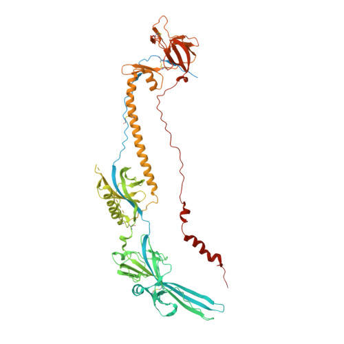
Members of the Herpesviridae, including the medically important alphaherpesvirus varicella-zoster virus (VZV), induce fusion of the virion envelope with cell membranes during entry, and between cells to form polykaryocytes in infected tissues. The conserved glycoproteins, gB, gH and gL, are the core functional proteins of the herpesvirus fusion complex. gB serves as the primary fusogen via its fusion loops, but functions for the remaining gB domains remain unexplained. As a pathway for biological discovery of domain function, our approach used structure-based analysis of the viral fusogen together with a neutralizing antibody. We report here a 2.8 Å cryogenic-electron microscopy structure of native gB recovered from VZV-infected cells, in complex with a human monoclonal antibody, 93k. This high-resolution structure guided targeted mutagenesis at the gB-93k interface, providing compelling evidence that a domain spatially distant from the gB fusion loops is critical for herpesvirus fusion, revealing a potential new target for antiviral therapies.
- Department of Pediatrics, Stanford University School of Medicine, Stanford, CA, 94305, USA. sloliver@stanford.edu.
Organizational Affiliation: