


Structure of an anti-PEG antibody reveals an open ring that captures highly flexible PEG polymers
Huckaby, J.T., Jacobs, T.M., Li, Z., Perna, R.J., Wang, A., Nicely, N.I., Lai, S.K.(2020) Commun Chem 3: 124
Experimental Data Snapshot
Starting Model: experimental
View more details
(2020) Commun Chem 3: 124
Entity ID: 1 | |||||
|---|---|---|---|---|---|
| Molecule | Chains | Sequence Length | Organism | Details | Image |
| 6-3 Fab heavy chain | A, C, E, G [auth H] | 228 | Mus musculus | Mutation(s): 0 | 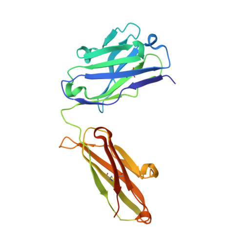 |
Entity ID: 2 | |||||
|---|---|---|---|---|---|
| Molecule | Chains | Sequence Length | Organism | Details | Image |
| 6-3 Fab light chain | B, D, F, H [auth L] | 219 | Mus musculus | Mutation(s): 0 | 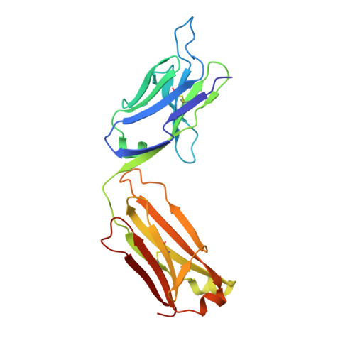 |
| Ligands 2 Unique | |||||
|---|---|---|---|---|---|
| ID | Chains | Name / Formula / InChI Key | 2D Diagram | 3D Interactions | |
| PEU (Subject of Investigation/LOI) Download:Ideal Coordinates CCD File | J [auth C], L [auth E] | 2,5,8,11,14,17,20,23,26,29,32,35,38,41,44,47,50,53,56,59,62,65,68,71,74,77,80-HEPTACOSAOXADOOCTACONTAN-82-OL C55 H112 O28 ISGUIIHZEJGUGQ-UHFFFAOYSA-N |  | ||
| EDO Download:Ideal Coordinates CCD File | I [auth A], K [auth D], M [auth H] | 1,2-ETHANEDIOL C2 H6 O2 LYCAIKOWRPUZTN-UHFFFAOYSA-N |  | ||
| Length ( Å ) | Angle ( ˚ ) |
|---|---|
| a = 112.108 | α = 90 |
| b = 87.791 | β = 113.655 |
| c = 116.074 | γ = 90 |
| Software Name | Purpose |
|---|---|
| PHENIX | refinement |
| HKL-2000 | data reduction |
| HKL-2000 | data scaling |
| PHASER | phasing |
| Funding Organization | Location | Grant Number |
|---|---|---|
| National Institutes of Health/National Institute of Dental and Craniofacial Research (NIH/NIDCR) | United States | F32DE026683 |
| National Science Foundation (NSF, United States) | United States | DMR-1810168 |
| National Science Foundation (NSF, United States) | United States | DGE-1650116 |
| David and Lucile Packard Foundation | United States | 2013-39274 |
| National Institutes of Health/National Heart, Lung, and Blood Institute (NIH/NHLBI) | United States | HL141934 |


