6VJP | pdb_00006vjp

Structure of Staphylococcus aureus peptidoglycan O-acetyltransferase A (OatA) C-terminal catalytic domain


Experimental Data Snapshot

  • Method: X-RAY DIFFRACTION
  • Resolution: 1.71 Å
  • R-Value Free: 
    0.195 (Depositor), 0.197 (DCC) 
  • R-Value Work: 
    0.166 (Depositor), 0.168 (DCC) 
  • R-Value Observed: 
    0.168 (Depositor) 

Starting Model: experimental
View more details

wwPDB Validation 3D Report Full Report

Validation slider image for 6VJP

This is version 1.4 of the entry. See complete history

Literature

Structural basis for theO-acetyltransferase function of the extracytoplasmic domain of OatA fromStaphylococcus aureus.

Jones, C.S.Sychantha, D.Howell, P.L.Clarke, A.J.

(2020) J Biological Chem 295: 8204-8213

  • DOI: https://doi.org/10.1074/jbc.RA120.013108
  • Primary Citation Related Structures: 
    6VJP, 6WN9

  • PubMed Abstract: 

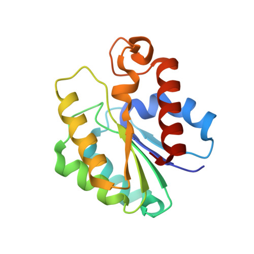
    Many bacteria possess enzymes that modify the essential cell-wall polymer peptidoglycan by O -acetylation. This modification occurs in numerous Gram-positive pathogens, including methicillin-resistant Staphylococcus aureus , a common cause of human infections. O -Acetylation of peptidoglycan protects bacteria from the lytic activity of lysozyme, a mammalian innate immune enzyme, and as such is important for bacterial virulence. The O -acetylating enzyme in Gram-positive bacteria, O -acetyltransferase A (OatA), is a two-domain protein consisting of an N-terminal integral membrane domain and a C-terminal extracytoplasmic domain. Here, we present the X-ray crystal structure at 1.71 Å resolution and the biochemical characterization of the C-terminal domain of S. aureus OatA. The structure revealed that this OatA domain adopts an SGNH-hydrolase fold and possesses a canonical catalytic triad. Site-specific replacement of active-site amino acids revealed the presence of a water-coordinating aspartate residue that limits esterase activity. This residue, although conserved in staphyloccocal OatA and most other homologs, is not present in the previously characterized streptococcal OatA. These results provide insights into the mechanism of acetyl transfer in the SGNH/GDSL hydrolase family and highlight important evolutionary differences between homologous OatA enzymes. Furthermore, this study enhances our understanding of PG O -acetyltransferases, which could guide the development of novel antibacterial drugs to combat infections with multidrug-resistant bacterial pathogens.


  • Organizational Affiliation
    • Department of Molecular and Cellular Biology, University of Guelph, Guelph, Ontario, Canada.

Macromolecule Content 

  • Total Structure Weight: 34.99 kDa 
  • Atom Count: 2,693 
  • Modeled Residue Count: 308 
  • Deposited Residue Count: 318 
  • Unique protein chains: 1

Macromolecules

Find similar proteins by:|  3D Structure
Entity ID: 1
MoleculeChains  Sequence LengthOrganismDetailsImage
Acetyltransferase
A, B
159Staphylococcus aureusMutation(s): 2 
Gene Names: E5491_14435
EC: 2.3.1
UniProt
Find proteins for Q2FV54 (Staphylococcus aureus (strain NCTC 8325 / PS 47))
Explore Q2FV54 
Go to UniProtKB:  Q2FV54
Entity Groups
Sequence Clusters30% Identity50% Identity70% Identity90% Identity95% Identity100% Identity
UniProt GroupQ2FV54
Sequence Annotations
Expand
Reference Sequence

Experimental Data & Validation

Experimental Data

  • Method: X-RAY DIFFRACTION
  • Resolution: 1.71 Å
  • R-Value Free:  0.195 (Depositor), 0.197 (DCC) 
  • R-Value Work:  0.166 (Depositor), 0.168 (DCC) 
  • R-Value Observed: 0.168 (Depositor) 
Space Group: P 1 21 1
Unit Cell:
Length ( Å )Angle ( ˚ )
a = 42.347α = 90
b = 61.303β = 100.9
c = 67.69γ = 90
Software Package:
Software NamePurpose
PHENIXrefinement
PDB_EXTRACTdata extraction
HKL-2000data reduction
HKL-2000data scaling
PHASERphasing

Structure Validation

View Full Validation Report


Entry History 

& Funding Information

Deposition Data


Funding OrganizationLocationGrant Number
Canadian Institutes of Health Research (CIHR)CanadaPJT 156353
Canadian Glycomics NetworkCanada--

Revision History  (Full details and data files)

  • Version 1.0: 2020-05-06
    Type: Initial release
  • Version 1.1: 2020-05-13
    Changes: Database references
  • Version 1.2: 2020-06-24
    Changes: Database references
  • Version 1.3: 2024-03-06
    Changes: Data collection, Database references, Derived calculations
  • Version 1.4: 2024-04-03
    Changes: Refinement description