6VJA

Structure of CD20 in complex with rituximab Fab


Experimental Data Snapshot

  • Method: ELECTRON MICROSCOPY
  • Resolution: 3.30 Å
  • Aggregation State: PARTICLE 
  • Reconstruction Method: SINGLE PARTICLE 

wwPDB Validation   3D Report Full Report


This is version 1.2 of the entry. See complete history


Literature

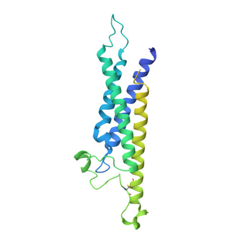
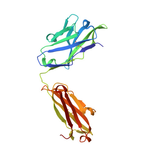
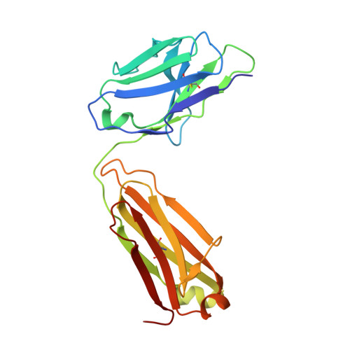
Structure of CD20 in complex with the therapeutic monoclonal antibody rituximab.

Rouge, L.Chiang, N.Steffek, M.Kugel, C.Croll, T.I.Tam, C.Estevez, A.Arthur, C.P.Koth, C.M.Ciferri, C.Kraft, E.Payandeh, J.Nakamura, G.Koerber, J.T.Rohou, A.

(2020) Science 367: 1224-1230

  • DOI: https://doi.org/10.1126/science.aaz9356
  • Primary Citation of Related Structures:  
    6VJA

  • PubMed Abstract: 

    Cluster of differentiation 20 (CD20) is a B cell membrane protein that is targeted by monoclonal antibodies for the treatment of malignancies and autoimmune disorders but whose structure and function are unknown. Rituximab (RTX) has been in clinical use for two decades, but how it activates complement to kill B cells remains poorly understood. We obtained a structure of CD20 in complex with RTX, revealing CD20 as a compact double-barrel dimer bound by two RTX antigen-binding fragments (Fabs), each of which engages a composite epitope and an extensive homotypic Fab:Fab interface. Our data suggest that RTX cross-links CD20 into circular assemblies and lead to a structural model for complement recruitment. Our results further highlight the potential relevance of homotypic Fab:Fab interactions in targeting oligomeric cell-surface markers.


  • Organizational Affiliation

    Department of Structural Biology, Genentech Inc., South San Francisco, CA 94080, USA.


Macromolecules
Find similar proteins by:  (by identity cutoff)  |  3D Structure
Entity ID: 1
MoleculeChains Sequence LengthOrganismDetailsImage
B-lymphocyte antigen CD20A [auth C],
B [auth D]
278Homo sapiensMutation(s): 0 
Gene Names: MS4A1CD20
Membrane Entity: Yes 
UniProt & NIH Common Fund Data Resources
Find proteins for P11836 (Homo sapiens)
Explore P11836 
Go to UniProtKB:  P11836
PHAROS:  P11836
GTEx:  ENSG00000156738 
Entity Groups  
Sequence Clusters30% Identity50% Identity70% Identity90% Identity95% Identity100% Identity
UniProt GroupP11836
Sequence Annotations
Expand
  • Reference Sequence
Find similar proteins by:  (by identity cutoff)  |  3D Structure
Entity ID: 2
MoleculeChains Sequence LengthOrganismDetailsImage
Rituximab Fab heavy chainC [auth H],
D [auth I]
224Homo sapiensMutation(s): 0 
Entity Groups  
Sequence Clusters30% Identity50% Identity70% Identity90% Identity95% Identity100% Identity
Sequence Annotations
Expand
  • Reference Sequence
Find similar proteins by:  (by identity cutoff)  |  3D Structure
Entity ID: 3
MoleculeChains Sequence LengthOrganismDetailsImage
Rituximab Fab light chainE [auth L],
F [auth M]
213Homo sapiensMutation(s): 0 
Entity Groups  
Sequence Clusters30% Identity50% Identity70% Identity90% Identity95% Identity100% Identity
Sequence Annotations
Expand
  • Reference Sequence
Experimental Data & Validation

Experimental Data

  • Method: ELECTRON MICROSCOPY
  • Resolution: 3.30 Å
  • Aggregation State: PARTICLE 
  • Reconstruction Method: SINGLE PARTICLE 
EM Software:
TaskSoftware PackageVersion
RECONSTRUCTIONcisTEM
MODEL REFINEMENTPHENIX

Structure Validation

View Full Validation Report


Entry History 

Deposition Data

Revision History  (Full details and data files)

  • Version 1.0: 2020-02-26
    Type: Initial release
  • Version 1.1: 2020-03-04
    Changes: Database references
  • Version 1.2: 2020-03-25
    Changes: Database references