6VD2 | pdb_00006vd2

Crystal structure of Arabidopsis thaliana S-adenosylmethionine Synthase 2 (AtMAT2) in complex with S-adenosylmethionine


Experimental Data Snapshot

  • Method: X-RAY DIFFRACTION
  • Resolution: 1.97 Å
  • R-Value Free: 
    0.240 (Depositor), 0.259 (DCC) 
  • R-Value Work: 
    0.191 (Depositor), 0.208 (DCC) 
  • R-Value Observed: 
    0.192 (Depositor) 

Starting Model: experimental
View more details

wwPDB Validation 3D Report Full Report

Validation slider image for 6VD2

Ligand Structure Quality Assessment 


This is version 1.2 of the entry. See complete history

Literature

S-adenosylmethionine synthases in plants: Structural characterization of type I and II isoenzymes from Arabidopsis thaliana and Medicago truncatula.

Sekula, B.Ruszkowski, M.Dauter, Z.

(2020) Int J Biol Macromol 151: 554-565

  • DOI: https://doi.org/10.1016/j.ijbiomac.2020.02.100
  • Primary Citation Related Structures: 
    6VCW, 6VCX, 6VCY, 6VCZ, 6VD0, 6VD1, 6VD2

  • PubMed Abstract: 

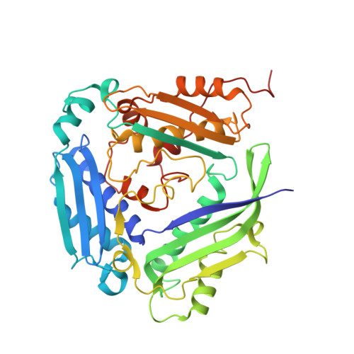
    S-adenosylmethionine synthases (MATs) are responsible for production of S-adenosylmethionine, the cofactor essential for various methylation reactions, production of polyamines and phytohormone ethylene, etc. Plants have two distinct MAT types (I and II). This work presents the structural analysis of MATs from Arabidopsis thaliana (AtMAT1 and AtMAT2, both type I) and Medicago truncatula (MtMAT3a, type II), which, unlike most MATs from other domains of life, are dimers where three-domain subunits are sandwiched flat with one another. Although MAT types are very similar, their subunits are differently oriented within the dimer. Structural snapshots along the enzymatic reaction reveal the exact conformation of precatalytic methionine in the active site and show a binding niche, characteristic only for plant MATs, that may serve as a lock of the gate loop. Nevertheless, plants, in contrary to mammals, lack the MAT regulatory subunit, and the regulation of plant MAT activity is still puzzling. Our structures open a possibility of an allosteric activity regulation of type I plant MATs by linear compounds, like polyamines, which would tighten the relationship between S-adenosylmethionine and polyamine biosynthesis.


  • Organizational Affiliation
    • Synchrotron Radiation Research Section, Macromolecular Crystallography Laboratory, National Cancer Institute, Argonne, IL, USA. Electronic address: bartosz.sekula@nih.gov.

Macromolecule Content 

  • Total Structure Weight: 90.94 kDa 
  • Atom Count: 6,882 
  • Modeled Residue Count: 786 
  • Deposited Residue Count: 804 
  • Unique protein chains: 1

Macromolecules

Find similar proteins by:|  3D Structure
Entity ID: 1
MoleculeChains  Sequence LengthOrganismDetailsImage
S-adenosylmethionine synthase 2
A, B
402Arabidopsis thalianaMutation(s): 0 
Gene Names: SAM2At4g01850T7B11.11
EC: 2.5.1.6
UniProt
Find proteins for P17562 (Arabidopsis thaliana)
Explore P17562 
Go to UniProtKB:  P17562
Entity Groups
Sequence Clusters30% Identity50% Identity70% Identity90% Identity95% Identity100% Identity
UniProt GroupP17562
Sequence Annotations
Expand
Reference Sequence

Small Molecules

Ligands 8 Unique
IDChains Name / Formula / InChI Key2D Diagram3D Interactions
SAM
(Subject of Investigation/LOI)

Query on SAM


Download:Ideal Coordinates CCD File
C [auth A],
N [auth B]
S-ADENOSYLMETHIONINE
C15 H22 N6 O5 S
MEFKEPWMEQBLKI-FCKMPRQPSA-N
PE8

Query on PE8


Download:Ideal Coordinates CCD File
M [auth A]3,6,9,12,15,18,21-HEPTAOXATRICOSANE-1,23-DIOL
C16 H34 O9
GLZWNFNQMJAZGY-UHFFFAOYSA-N
MPO

Query on MPO


Download:Ideal Coordinates CCD File
K [auth A],
T [auth B]
3[N-MORPHOLINO]PROPANE SULFONIC ACID
C7 H15 N O4 S
DVLFYONBTKHTER-UHFFFAOYSA-N
PGE

Query on PGE


Download:Ideal Coordinates CCD File
J [auth A]TRIETHYLENE GLYCOL
C6 H14 O4
ZIBGPFATKBEMQZ-UHFFFAOYSA-N
PEG

Query on PEG


Download:Ideal Coordinates CCD File
L [auth A],
U [auth B]
DI(HYDROXYETHYL)ETHER
C4 H10 O3
MTHSVFCYNBDYFN-UHFFFAOYSA-N
PO4

Query on PO4


Download:Ideal Coordinates CCD File
G [auth A],
H [auth A],
Q [auth B],
R [auth B],
S [auth B]
PHOSPHATE ION
O4 P
NBIIXXVUZAFLBC-UHFFFAOYSA-K
K
(Subject of Investigation/LOI)

Query on K


Download:Ideal Coordinates CCD File
F [auth A],
P [auth B]
POTASSIUM ION
K
NPYPAHLBTDXSSS-UHFFFAOYSA-N
MG
(Subject of Investigation/LOI)

Query on MG


Download:Ideal Coordinates CCD File
D [auth A],
E [auth A],
I [auth A],
O [auth B]
MAGNESIUM ION
Mg
JLVVSXFLKOJNIY-UHFFFAOYSA-N

Experimental Data & Validation

Experimental Data

  • Method: X-RAY DIFFRACTION
  • Resolution: 1.97 Å
  • R-Value Free:  0.240 (Depositor), 0.259 (DCC) 
  • R-Value Work:  0.191 (Depositor), 0.208 (DCC) 
  • R-Value Observed: 0.192 (Depositor) 
Space Group: P 1 21 1
Unit Cell:
Length ( Å )Angle ( ˚ )
a = 61.98α = 90
b = 101.354β = 99.8
c = 84.708γ = 90
Software Package:
Software NamePurpose
REFMACrefinement
PDB_EXTRACTdata extraction
XDSdata reduction
XSCALEdata scaling
PHASERphasing

Structure Validation

View Full Validation Report


Ligand Structure Quality Assessment 


Entry History 

& Funding Information

Deposition Data


Funding OrganizationLocationGrant Number
National Institutes of Health/National Cancer Institute (NIH/NCI)United StatesThe Intramural Research Program

Revision History  (Full details and data files)

  • Version 1.0: 2020-02-26
    Type: Initial release
  • Version 1.1: 2020-03-11
    Changes: Database references
  • Version 1.2: 2023-10-11
    Changes: Data collection, Database references, Derived calculations, Refinement description