6VCL

Crystal structure of E.coli RppH-DapF in complex with pppGpp, Mg2+ and F-


Experimental Data Snapshot

  • Method: X-RAY DIFFRACTION
  • Resolution: 2.06 Å
  • R-Value Free: 0.227 
  • R-Value Work: 0.191 
  • R-Value Observed: 0.193 

wwPDB Validation   3D Report Full Report

Currently 6VCL does not have a validation slider image.


This is version 1.2 of the entry. See complete history


Literature

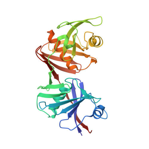
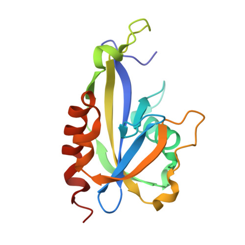
Principles of RNA and nucleotide discrimination by the RNA processing enzyme RppH.

Gao, A.Vasilyev, N.Kaushik, A.Duan, W.Serganov, A.

(2020) Nucleic Acids Res 48: 3776-3788

  • DOI: https://doi.org/10.1093/nar/gkaa024
  • Primary Citation of Related Structures:  
    6VCK, 6VCL, 6VCM, 6VCN, 6VCO, 6VCP, 6VCQ, 6VCR

  • PubMed Abstract: 

    All enzymes face a challenge of discriminating cognate substrates from similar cellular compounds. Finding a correct substrate is especially difficult for the Escherichia coli Nudix hydrolase RppH, which triggers 5'-end-dependent RNA degradation by removing orthophosphate from the 5'-diphosphorylated transcripts. Here we show that RppH binds and slowly hydrolyzes NTPs, NDPs and (p)ppGpp, which each resemble the 5'-end of RNA. A series of X-ray crystal structures of RppH-nucleotide complexes, trapped in conformations either compatible or incompatible with hydrolysis, explain the low reaction rates of mononucleotides and suggest two distinct mechanisms for their hydrolysis. While RppH adopts the same catalytic arrangement with 5'-diphosphorylated nucleotides as with RNA, the enzyme hydrolyzes 5'-triphosphorylated nucleotides by extending the active site with an additional Mg2+ cation, which coordinates another reactive nucleophile. Although the average intracellular pH minimizes the hydrolysis of nucleotides by slowing their reaction with RppH, they nevertheless compete with RNA for binding and differentially inhibit the reactivity of RppH with triphosphorylated and diphosphorylated RNAs. Thus, E. coli RppH integrates various signals, such as competing non-cognate substrates and a stimulatory protein factor DapF, to achieve the differential degradation of transcripts involved in cellular processes important for the adaptation of bacteria to different growth conditions.


  • Organizational Affiliation

    Department of Biochemistry and Molecular Pharmacology, New York University School of Medicine, 550 First Avenue, New York, NY 10016, USA.


Macromolecules
Find similar proteins by:  (by identity cutoff)  |  3D Structure
Entity ID: 1
MoleculeChains Sequence LengthOrganismDetailsImage
Diaminopimelate epimerase275Escherichia coli K-12Mutation(s): 2 
Gene Names: dapFb3809JW5592
EC: 5.1.1.7
UniProt
Find proteins for P0A6K1 (Escherichia coli (strain K12))
Explore P0A6K1 
Go to UniProtKB:  P0A6K1
Entity Groups  
Sequence Clusters30% Identity50% Identity70% Identity90% Identity95% Identity100% Identity
UniProt GroupP0A6K1
Sequence Annotations
Expand
  • Reference Sequence
Find similar proteins by:  (by identity cutoff)  |  3D Structure
Entity ID: 2
MoleculeChains Sequence LengthOrganismDetailsImage
RNA pyrophosphohydrolase161Escherichia coli K-12Mutation(s): 2 
Gene Names: rppHnudHygdPb2830JW2798
EC: 3.6.1
UniProt
Find proteins for P0A776 (Escherichia coli (strain K12))
Explore P0A776 
Go to UniProtKB:  P0A776
Entity Groups  
Sequence Clusters30% Identity50% Identity70% Identity90% Identity95% Identity100% Identity
UniProt GroupP0A776
Sequence Annotations
Expand
  • Reference Sequence
Small Molecules
Ligands 5 Unique
IDChains Name / Formula / InChI Key2D Diagram3D Interactions
0O2 (Subject of Investigation/LOI)
Query on 0O2

Download Ideal Coordinates CCD File 
L [auth B]guanosine 5'-(tetrahydrogen triphosphate) 3'-(trihydrogen diphosphate)
C10 H18 N5 O20 P5
KCPMACXZAITQAX-UUOKFMHZSA-N
GOL
Query on GOL

Download Ideal Coordinates CCD File 
H [auth B],
I [auth B]
GLYCEROL
C3 H8 O3
PEDCQBHIVMGVHV-UHFFFAOYSA-N
CL
Query on CL

Download Ideal Coordinates CCD File 
C [auth A],
J [auth B]
CHLORIDE ION
Cl
VEXZGXHMUGYJMC-UHFFFAOYSA-M
MG (Subject of Investigation/LOI)
Query on MG

Download Ideal Coordinates CCD File 
D [auth B],
E [auth B],
F [auth B],
G [auth B]
MAGNESIUM ION
Mg
JLVVSXFLKOJNIY-UHFFFAOYSA-N
F (Subject of Investigation/LOI)
Query on F

Download Ideal Coordinates CCD File 
K [auth B]FLUORIDE ION
F
KRHYYFGTRYWZRS-UHFFFAOYSA-M
Binding Affinity Annotations 
IDSourceBinding Affinity
0O2 Binding MOAD:  6VCL IC50: 5.44e+4 (nM) from 1 assay(s)
Experimental Data & Validation

Experimental Data

  • Method: X-RAY DIFFRACTION
  • Resolution: 2.06 Å
  • R-Value Free: 0.227 
  • R-Value Work: 0.191 
  • R-Value Observed: 0.193 
  • Space Group: C 2 2 21
Unit Cell:
Length ( Å )Angle ( ˚ )
a = 162.444α = 90
b = 190.649β = 90
c = 51.336γ = 90
Software Package:
Software NamePurpose
PHENIXrefinement
HKL-2000data reduction
HKL-2000data scaling
Cootmodel building
PHENIXphasing

Structure Validation

View Full Validation Report

Currently 6VCL does not have a validation slider image.


Entry History & Funding Information

Deposition Data


Funding OrganizationLocationGrant Number
National Institutes of Health/National Human Genome Research Institute (NIH/NHGRI)United StatesR01GM112940

Revision History  (Full details and data files)

  • Version 1.0: 2020-02-05
    Type: Initial release
  • Version 1.1: 2020-04-22
    Changes: Database references
  • Version 1.2: 2023-10-11
    Changes: Data collection, Database references, Derived calculations, Refinement description