6VCA | pdb_00006vca

TB38 complex


Experimental Data Snapshot

  • Method: X-RAY DIFFRACTION
  • Resolution: 3.73 Å
  • R-Value Free: 
    0.264 (Depositor) 
  • R-Value Work: 
    0.224 (Depositor) 
  • R-Value Observed: 
    0.226 (Depositor) 

Starting Model: experimental
View more details

wwPDB Validation 3D Report Full Report

Validation slider image for 6VCA

Ligand Structure Quality Assessment 


This is version 1.3 of the entry. See complete history

Literature

A highly potent CD73 biparatopic antibody blocks organization of the enzyme active site through dual mechanisms.

Stefano, J.E.Lord, D.M.Zhou, Y.Jaworski, J.Hopke, J.Travaline, T.Zhang, N.Wong, K.Lennon, A.He, T.Bric-Furlong, E.Cherrie, C.Magnay, T.Remy, E.Brondyk, W.Qiu, H.Radosevic, K.

(2020) J Biological Chem 295: 18379-18389

  • DOI: https://doi.org/10.1074/jbc.RA120.012395
  • Primary Citation Related Structures: 
    6VC9, 6VCA

  • PubMed Abstract: 

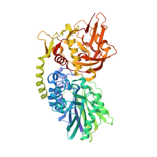
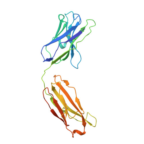
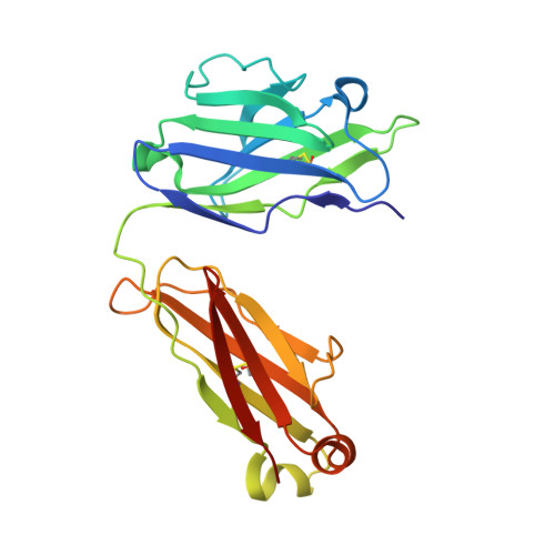
    The dimeric ectonucleotidase CD73 catalyzes the hydrolysis of AMP at the cell surface to form adenosine, a potent suppressor of the immune response. Blocking CD73 activity in the tumor microenvironment can have a beneficial effect on tumor eradication and is a promising approach for cancer therapy. Biparatopic antibodies binding different regions of CD73 may be a means to antagonize its enzymatic activity. A panel of biparatopic antibodies representing the pairwise combination of 11 parental monoclonal antibodies against CD73 was generated by Fab-arm exchange. Nine variants vastly exceeded the potency of their parental antibodies with ≥90% inhibition of activity and subnanomolar EC 50 values. Pairing the Fabs of parents with nonoverlapping epitopes was both sufficient and necessary whereas monovalent antibodies were poor inhibitors. Some parental antibodies yielded potent biparatopics with multiple partners, one of which (TB19) producing the most potent. The structure of the TB19 Fab with CD73 reveals that it blocks alignment of the N- and C-terminal CD73 domains necessary for catalysis. A separate structure of CD73 with a Fab (TB38) which complements TB19 in a particularly potent biparatopic shows its binding to a nonoverlapping site on the CD73 N-terminal domain. Structural modeling demonstrates a TB19/TB38 biparatopic antibody would be unable to bind the CD73 dimer in a bivalent manner, implicating crosslinking of separate CD73 dimers in its mechanism of action. This ability of a biparatopic antibody to both crosslink CD73 dimers and fix them in an inactive conformation thus represents a highly effective mechanism for the inhibition of CD73 activity.


  • Organizational Affiliation
    • Biologics Research, Sanofi R&D Framingham, Massachusetts, USA.

Macromolecule Content 

  • Total Structure Weight: 428.81 kDa 
  • Atom Count: 27,834 
  • Modeled Residue Count: 3,616 
  • Deposited Residue Count: 3,912 
  • Unique protein chains: 3

Macromolecules

Find similar proteins by:|  3D Structure
Entity ID: 1
MoleculeChains  Sequence LengthOrganismDetailsImage
5'-nucleotidase, ecto (CD73), isoform CRA_a
A, B, C, D
529Homo sapiensMutation(s): 0 
Gene Names: NT5EhCG_401111
EC: 3.1.3.5 (PDB Primary Data), 3.1.3.99 (UniProt), 3.1.3.89 (UniProt), 3.1.3.35 (UniProt), 3.1.3.91 (UniProt)
UniProt & NIH Common Fund Data Resources
Find proteins for P21589 (Homo sapiens)
Explore P21589 
Go to UniProtKB:  P21589
PHAROS:  P21589
GTEx:  ENSG00000135318 
Entity Groups
Sequence Clusters30% Identity50% Identity70% Identity90% Identity95% Identity100% Identity
UniProt GroupP21589
Glycosylation
Glycosylation Sites: 1Go to GlyGen: P21589-1
Sequence Annotations
Expand
Reference Sequence
Find similar proteins by:|  3D Structure
Entity ID: 2
MoleculeChains  Sequence LengthOrganismDetailsImage
TB38 heavy chainE [auth H],
G [auth E],
I [auth P],
K
233Homo sapiensMutation(s): 0 
Find similar proteins by:|  3D Structure
Entity ID: 3
MoleculeChains  Sequence LengthOrganismDetailsImage
TB38 light chainF [auth L],
H [auth F],
J [auth O],
L [auth J]
216Homo sapiensMutation(s): 0 

Oligosaccharides

Help  
Entity ID: 4
MoleculeChains Length2D Diagram GlycosylationD Interactions
2-acetamido-2-deoxy-beta-D-glucopyranose-(1-4)-2-acetamido-2-deoxy-beta-D-glucopyranoseM [auth G]2N-Glycosylation
Glycosylation Resources
GlyTouCan: G42666HT
GlyCosmos: G42666HT
GlyGen: G42666HT
Entity ID: 5
MoleculeChains Length2D Diagram GlycosylationD Interactions
beta-D-mannopyranose-(1-4)-2-acetamido-2-deoxy-beta-D-glucopyranose-(1-4)-2-acetamido-2-deoxy-beta-D-glucopyranoseN [auth I]3N-Glycosylation
Glycosylation Resources
GlyTouCan: G15407YE
GlyCosmos: G15407YE
GlyGen: G15407YE

Small Molecules

Ligands 1 Unique
IDChains Name / Formula / InChI Key2D Diagram3D Interactions
NAG

Query on NAG


Download:Ideal Coordinates CCD File
O [auth D]2-acetamido-2-deoxy-beta-D-glucopyranose
C8 H15 N O6
OVRNDRQMDRJTHS-FMDGEEDCSA-N

Experimental Data & Validation

Experimental Data

  • Method: X-RAY DIFFRACTION
  • Resolution: 3.73 Å
  • R-Value Free:  0.264 (Depositor) 
  • R-Value Work:  0.224 (Depositor) 
  • R-Value Observed: 0.226 (Depositor) 
Space Group: C 2 2 2
Unit Cell:
Length ( Å )Angle ( ˚ )
a = 236.91α = 90
b = 336.2β = 90
c = 222.15γ = 90
Software Package:
Software NamePurpose
XSCALEdata scaling
REFMACrefinement
PDB_EXTRACTdata extraction
XDSdata reduction
MOLREPphasing

Structure Validation

View Full Validation Report


Ligand Structure Quality Assessment 


Entry History 

Deposition Data

Revision History  (Full details and data files)

  • Version 1.0: 2020-11-11
    Type: Initial release
  • Version 1.1: 2021-01-06
    Changes: Database references
  • Version 1.2: 2023-10-11
    Changes: Data collection, Database references, Refinement description
  • Version 1.3: 2024-10-30
    Changes: Structure summary