Structure-Guided Engineering of the Homodimeric Mango-IV Fluorescence Turn-on Aptamer Yields an RNA FRET Pair.
Trachman 3rd, R.J., Cojocaru, R., Wu, D., Piszczek, G., Ryckelynck, M., Unrau, P.J., Ferre-D'Amare, A.R.(2020) Structure 28: 776-785.e3
- PubMed: 32386573 Search on PubMedSearch on PubMed Central
- DOI: https://doi.org/10.1016/j.str.2020.04.007
- Primary Citation Related Structures:
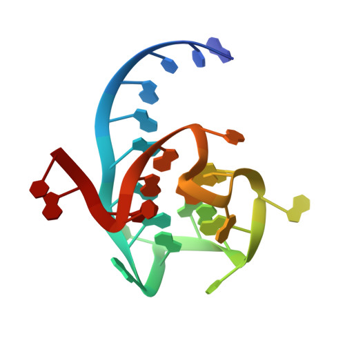
6V9B, 6V9D - PubMed Abstract:
Fluorescent RNA aptamers have been used in cells as biosensor reporters and tags for tracking transcripts. Recently, combined SELEX and microfluidic fluorescence sorting yielded three aptamers that activate fluorescence of TO1-Biotin: Mango-II, Mango-III, and Mango-IV. Of these, Mango-IV was best at imaging RNAs in both fixed and live mammalian cells. To understand how Mango-IV achieves activity in cells, we determined its crystal structure complexed with TO1-Biotin. The structure reveals a domain-swapped homodimer with two independent G-quadruplex fluorophore binding pockets. Structure-based analyses indicate that the Mango-IV core has relaxed fluorophore specificity, and a tendency to reorganize binding pocket residues. These molecular properties may endow it with robustness in the cellular milieu. Based on the domain-swapped structure, heterodimers between Mango-IV and the fluorescent aptamer iSpinach, joined by Watson-Crick base pairing, were constructed. These exhibited FRET between their respective aptamer-activated fluorophores, advancing fluorescent aptamer technology toward multi-color, RNA-based imaging of RNA coexpression and colocalization.
- Biochemistry and Biophysics Center, National Heart, Lung, and Blood Institute, 50 South Drive MSC 8012, Bethesda, MD 20892-8012, USA.
Organizational Affiliation: