6V8A

Human CtBP1 (28-375) in complex with AMP

  • Classification: TRANSCRIPTION
  • Organism(s): Homo sapiens
  • Expression System: Escherichia coli
  • Mutation(s): Yes 

  • Deposited: 2019-12-10 Released: 2021-02-03 
  • Deposition Author(s): Royer, W.E.
  • Funding Organization(s): National Institutes of Health/National Institute of General Medical Sciences (NIH/NIGMS)

Experimental Data Snapshot

  • Method: X-RAY DIFFRACTION
  • Resolution: 2.35 Å
  • R-Value Free: 0.258 
  • R-Value Work: 0.203 
  • R-Value Observed: 0.205 

wwPDB Validation   3D Report Full Report

Currently 6V8A does not have a validation slider image.


This is version 1.3 of the entry. See complete history


Literature

NAD(H) phosphates mediate tetramer assembly of human C-terminal binding protein (CtBP).

Nichols, J.C.Schiffer, C.A.Royer Jr., W.E.

(2021) J Biol Chem 296: 100351-100351

  • DOI: https://doi.org/10.1016/j.jbc.2021.100351
  • Primary Citation of Related Structures:  
    6V89, 6V8A, 7KWM

  • PubMed Abstract: 

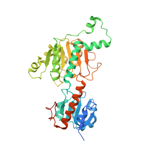
    C-terminal binding proteins (CtBPs) are cotranscriptional factors that play key roles in cell fate. We have previously shown that NAD(H) promotes the assembly of similar tetramers from either human CtBP1 and CtBP2 and that CtBP2 tetramer destabilizing mutants are defective for oncogenic activity. To assist structure-based design efforts for compounds that disrupt CtBP tetramerization, it is essential to understand how NAD(H) triggers tetramer assembly. Here, we investigate the moieties within NAD(H) that are responsible for triggering tetramer formation. Using multiangle light scattering (MALS), we show that ADP is able to promote tetramer formation of both CtBP1 and CtBP2, whereas AMP promotes tetramer assembly of CtBP1, but not CtBP2. Other NAD(H) moieties that lack the adenosine phosphate, including adenosine and those incorporating nicotinamide, all fail to promote tetramer assembly. Our crystal structures of CtBP1 with AMP reveal participation of the adenosine phosphate in the tetrameric interface, pinpointing its central role in NAD(H)-linked assembly. CtBP1 and CtBP2 have overlapping but unique roles, suggesting that a detailed understanding of their unique structural properties might have utility in the design of paralog-specific inhibitors. We investigated the different responses to AMP through a series of site-directed mutants at 13 positions. These mutations reveal a central role for a hinge segment, which we term the 120s hinge that connects the substrate with coenzyme-binding domains and influences nucleotide binding and tetramer assembly. Our results provide insight into suitable pockets to explore in structure-based drug design to interfere with cotranscriptional activity of CtBP in cancer.


  • Organizational Affiliation

    Department of Biochemistry and Molecular Pharmacology, University of Massachusetts Medical School, Worcester, Massachusetts, USA; Chemistry Department, Worcester State University, Worcester, Massachusetts, USA.


Macromolecules
Find similar proteins by:  (by identity cutoff)  |  3D Structure
Entity ID: 1
MoleculeChains Sequence LengthOrganismDetailsImage
C-terminal-binding protein 1369Homo sapiensMutation(s): 1 
Gene Names: CTBP1CTBP
EC: 1.1.1
UniProt & NIH Common Fund Data Resources
Find proteins for Q13363 (Homo sapiens)
Explore Q13363 
Go to UniProtKB:  Q13363
PHAROS:  Q13363
GTEx:  ENSG00000159692 
Entity Groups  
Sequence Clusters30% Identity50% Identity70% Identity90% Identity95% Identity100% Identity
UniProt GroupQ13363
Sequence Annotations
Expand
  • Reference Sequence
Experimental Data & Validation

Experimental Data

  • Method: X-RAY DIFFRACTION
  • Resolution: 2.35 Å
  • R-Value Free: 0.258 
  • R-Value Work: 0.203 
  • R-Value Observed: 0.205 
  • Space Group: P 64 2 2
Unit Cell:
Length ( Å )Angle ( ˚ )
a = 89.171α = 90
b = 89.171β = 90
c = 164.17γ = 120
Software Package:
Software NamePurpose
HKL-2000data scaling
PHENIXrefinement
PDB_EXTRACTdata extraction
HKL-3000data reduction
PHASERphasing

Structure Validation

View Full Validation Report

Currently 6V8A does not have a validation slider image.


Entry History & Funding Information

Deposition Data

  • Released Date: 2021-02-03 
  • Deposition Author(s): Royer, W.E.

Funding OrganizationLocationGrant Number
National Institutes of Health/National Institute of General Medical Sciences (NIH/NIGMS)United StatesR01 GM119014

Revision History  (Full details and data files)

  • Version 1.0: 2021-02-03
    Type: Initial release
  • Version 1.1: 2021-02-17
    Changes: Database references
  • Version 1.2: 2021-07-21
    Changes: Database references
  • Version 1.3: 2023-10-11
    Changes: Advisory, Data collection, Database references, Refinement description