6V7O | pdb_00006v7o

Structural Elucidation of Peptide Binding to KLHL-12, a Substrate Specific Adapter Protein in a Cul3-Ring E3 Ligase Complex

  • Classification: LIGASE
  • Organism(s): Homo sapiens
  • Expression System: Escherichia coli
  • Mutation(s): No 

  • Deposited: 2019-12-09 Released: 2020-02-19 
  • Deposition Author(s): Zhao, B.
  • Funding Organization(s): National Institutes of Health/National Cancer Institute (NIH/NCI)

Experimental Data Snapshot

  • Method: X-RAY DIFFRACTION
  • Resolution: 2.90 Å
  • R-Value Free: 
    0.254 (Depositor), 0.236 (DCC) 
  • R-Value Work: 
    0.207 (Depositor) 
  • R-Value Observed: 
    0.212 (Depositor) 

Starting Model: experimental
View more details

wwPDB Validation 3D Report Full Report

Validation slider image for 6V7O

This is version 1.2 of the entry. See complete history

Literature

Structural Elucidation of Peptide Binding to KLHL-12, a Substrate Specific Adapter Protein in a Cul3-Ring E3 Ligase Complex.

Zhao, B.Payne, W.G.Sai, J.Lu, Z.Olejniczak, E.T.Fesik, S.W.

(2020) Biochemistry 59: 964-969

  • DOI: https://doi.org/10.1021/acs.biochem.9b01073
  • Primary Citation Related Structures: 
    6V7O

  • PubMed Abstract: 

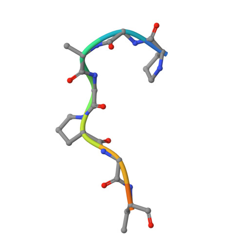
    KLHL-12 is a substrate specific adapter protein for a Cul3-Ring ligase complex. It is a member of the Kelch β-propeller domain subclass of Cullin-Ring substrate recognition domains. This E3 ubiquitin ligase complex has many activities, including acting as a negative regulator of the Wnt signaling pathway by mediating ubiquitination and subsequent proteolysis of Dvl3/Dsh3. KLHL-12 is also known to mediate the polyubiquitination of the dopamine D4 receptor (D4.2), the ubiquitination of KHSRP, a protein that is involved in IRES translation, and also the ubiquitination of Sec31, which is involved in endoplasmic reticulum-Golgi transport by regulating the size of COPII coats. Earlier studies broadly defined the substrate binding regions for D4.2 and Dvl3/Dsh3 to KLHL-12. We tested several peptides from these regions and succeeded in identifying a short peptide that bound to KLHL-12 with low micromolar affinity. To better understand the sequence specificity of this peptide, we used alanine substitutions to map the important residues and obtained an X-ray structure of this peptide bound to KLHL-12. This structure and our peptide affinity measurements suggest a sequence motif for peptides that bind to the top face of KLHL-12. Understanding this binding site on KLHL-12 may contribute to efforts to find small molecule ligands that can either directly inhibit the degradation of substrate proteins or be used in targeted protein degradation strategies using PROTACs.


  • Organizational Affiliation
    • Department of Biochemistry, Vanderbilt University School of Medicine, 2215 Garland Avenue, 607 Light Hall, Nashville, Tennessee 37232-0146, United States.

Macromolecule Content 

  • Total Structure Weight: 67.71 kDa 
  • Atom Count: 4,297 
  • Modeled Residue Count: 580 
  • Deposited Residue Count: 622 
  • Unique protein chains: 2

Macromolecules

Find similar proteins by:|  3D Structure
Entity ID: 1
MoleculeChains  Sequence LengthOrganismDetailsImage
Kelch-like protein 12
A, B
301Homo sapiensMutation(s): 0 
Gene Names: KLHL12C3IP1
UniProt & NIH Common Fund Data Resources
Find proteins for Q53G59 (Homo sapiens)
Explore Q53G59 
Go to UniProtKB:  Q53G59
PHAROS:  Q53G59
GTEx:  ENSG00000117153 
Entity Groups
Sequence Clusters30% Identity50% Identity70% Identity90% Identity95% Identity100% Identity
UniProt GroupQ53G59
Sequence Annotations
Expand
Reference Sequence
Find similar proteins by:  Sequence   |   3D Structure  
Entity ID: 2
MoleculeChains  Sequence LengthOrganismDetailsImage
Dvl3-peptide
C, D
10Homo sapiensMutation(s): 0 
UniProt & NIH Common Fund Data Resources
Find proteins for Q92997 (Homo sapiens)
Explore Q92997 
Go to UniProtKB:  Q92997
PHAROS:  Q92997
GTEx:  ENSG00000161202 
Entity Groups
Sequence Clusters30% Identity50% Identity70% Identity90% Identity95% Identity100% Identity
UniProt GroupQ92997
Sequence Annotations
Expand
Reference Sequence

Experimental Data & Validation

Experimental Data

  • Method: X-RAY DIFFRACTION
  • Resolution: 2.90 Å
  • R-Value Free:  0.254 (Depositor), 0.236 (DCC) 
  • R-Value Work:  0.207 (Depositor) 
  • R-Value Observed: 0.212 (Depositor) 
Space Group: P 1 21 1
Unit Cell:
Length ( Å )Angle ( ˚ )
a = 42.679α = 90
b = 105.124β = 90.18
c = 63.148γ = 90
Software Package:
Software NamePurpose
PHENIXrefinement
PDB_EXTRACTdata extraction
HKL-2000data reduction
HKL-2000data scaling
PHASERphasing

Structure Validation

View Full Validation Report


Entry History 

& Funding Information

Deposition Data

  • Released Date: 2020-02-19 
  • Deposition Author(s): Zhao, B.

Funding OrganizationLocationGrant Number
National Institutes of Health/National Cancer Institute (NIH/NCI)United States--

Revision History  (Full details and data files)

  • Version 1.0: 2020-02-19
    Type: Initial release
  • Version 1.1: 2020-03-18
    Changes: Database references
  • Version 1.2: 2023-10-11
    Changes: Data collection, Database references, Refinement description