6V55 | pdb_00006v55

Full extracellular region of zebrafish Gpr126/Adgrg6

  • Classification: CELL ADHESION
  • Organism(s): Danio rerio
  • Expression System: Trichoplusia ni
  • Mutation(s): No 

  • Deposited: 2019-12-03 Released: 2020-01-15 
  • Deposition Author(s): Leon, K., Arac, D.
  • Funding Organization(s): National Institutes of Health/National Institute of General Medical Sciences (NIH/NIGMS)

Experimental Data Snapshot

  • Method: X-RAY DIFFRACTION
  • Resolution: 2.38 Å
  • R-Value Free: 
    0.272 (Depositor), 0.271 (DCC) 
  • R-Value Work: 
    0.212 (Depositor), 0.225 (DCC) 
  • R-Value Observed: 
    0.215 (Depositor) 

wwPDB Validation 3D Report Full Report

Validation slider image for 6V55

This is version 1.3 of the entry. See complete history

Literature

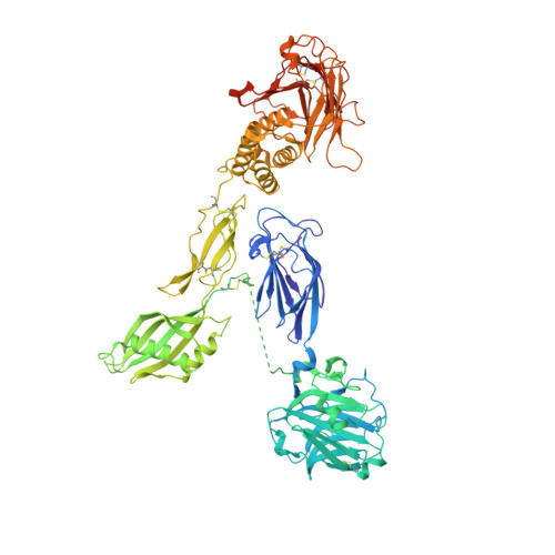
Structural basis for adhesion G protein-coupled receptor Gpr126 function.

Leon, K.Cunningham, R.L.Riback, J.A.Feldman, E.Li, J.Sosnick, T.R.Zhao, M.Monk, K.R.Arac, D.

(2020) Nat Commun 11: 194-194

  • DOI: https://doi.org/10.1038/s41467-019-14040-1
  • Primary Citation Related Structures: 
    6V55

  • PubMed Abstract: 

    Many drugs target the extracellular regions (ECRs) of cell-surface receptors. The large and alternatively-spliced ECRs of adhesion G protein-coupled receptors (aGPCRs) have key functions in diverse biological processes including neurodevelopment, embryogenesis, and tumorigenesis. However, their structures and mechanisms of action remain unclear, hampering drug development. The aGPCR Gpr126/Adgrg6 regulates Schwann cell myelination, ear canal formation, and heart development; and GPR126 mutations cause myelination defects in human. Here, we determine the structure of the complete zebrafish Gpr126 ECR and reveal five domains including a previously unknown domain. Strikingly, the Gpr126 ECR adopts a closed conformation that is stabilized by an alternatively spliced linker and a conserved calcium-binding site. Alternative splicing regulates ECR conformation and receptor signaling, while mutagenesis of the calcium-binding site abolishes Gpr126 function in vivo. These results demonstrate that Gpr126 ECR utilizes a multi-faceted dynamic approach to regulate receptor function and provide relevant insights for ECR-targeted drug design.


  • Organizational Affiliation
    • Department of Biochemistry and Molecular Biology, The University of Chicago, Chicago, IL, 60637, USA.

Macromolecule Content 

  • Total Structure Weight: 90.26 kDa 
  • Atom Count: 6,114 
  • Modeled Residue Count: 753 
  • Deposited Residue Count: 800 
  • Unique protein chains: 2

Macromolecules

Find similar proteins by:|  3D Structure
Entity ID: 1
MoleculeChains  Sequence LengthOrganismDetailsImage
Adhesion G-protein coupled receptor G6788Danio rerioMutation(s): 0 
Gene Names: adgrg6gpr126
UniProt
Find proteins for C6KFA3 (Danio rerio)
Explore C6KFA3 
Go to UniProtKB:  C6KFA3
Entity Groups
Sequence Clusters30% Identity50% Identity70% Identity90% Identity95% Identity100% Identity
UniProt GroupC6KFA3
Glycosylation
Glycosylation Sites: 11
Sequence Annotations
Expand
Reference Sequence
Find similar proteins by:  Sequence   |   3D Structure  
Entity ID: 2
MoleculeChains  Sequence LengthOrganismDetailsImage
Adhesion G-protein coupled receptor G6B [auth Q]12Danio rerioMutation(s): 0 
Gene Names: adgrg6gpr126
UniProt
Find proteins for C6KFA3 (Danio rerio)
Explore C6KFA3 
Go to UniProtKB:  C6KFA3
Entity Groups
Sequence Clusters30% Identity50% Identity70% Identity90% Identity95% Identity100% Identity
UniProt GroupC6KFA3
Sequence Annotations
Expand
Reference Sequence

Small Molecules

Ligands 2 Unique
IDChains Name / Formula / InChI Key2D Diagram3D Interactions
NAG

Query on NAG


Download:Ideal Coordinates CCD File
D [auth A]
E [auth A]
F [auth A]
G [auth A]
H [auth A]
D [auth A],
E [auth A],
F [auth A],
G [auth A],
H [auth A],
I [auth A],
J [auth A],
K [auth A],
L [auth A],
M [auth A],
N [auth A]
2-acetamido-2-deoxy-beta-D-glucopyranose
C8 H15 N O6
OVRNDRQMDRJTHS-FMDGEEDCSA-N
CA
(Subject of Investigation/LOI)

Query on CA


Download:Ideal Coordinates CCD File
C [auth A]CALCIUM ION
Ca
BHPQYMZQTOCNFJ-UHFFFAOYSA-N

Experimental Data & Validation

Experimental Data

  • Method: X-RAY DIFFRACTION
  • Resolution: 2.38 Å
  • R-Value Free:  0.272 (Depositor), 0.271 (DCC) 
  • R-Value Work:  0.212 (Depositor), 0.225 (DCC) 
  • R-Value Observed: 0.215 (Depositor) 
Space Group: C 1 2 1
Unit Cell:
Length ( Å )Angle ( ˚ )
a = 144.96α = 90
b = 59.448β = 107.821
c = 168.385γ = 90
Software Package:
Software NamePurpose
REFMACrefinement
PHENIXrefinement
HKL-2000data reduction
HKL-2000data scaling
CRANK2phasing

Structure Validation

View Full Validation Report


Entry History 

& Funding Information

Deposition Data


Funding OrganizationLocationGrant Number
National Institutes of Health/National Institute of General Medical Sciences (NIH/NIGMS)United StatesGM120322

Revision History  (Full details and data files)

  • Version 1.0: 2020-01-15
    Type: Initial release
  • Version 1.1: 2020-01-22
    Changes: Database references
  • Version 1.2: 2020-07-29
    Type: Remediation
    Reason: Carbohydrate remediation
    Changes: Data collection, Derived calculations, Structure summary
  • Version 1.3: 2024-10-09
    Changes: Data collection, Database references, Structure summary