6V4K

Structure of TrkH-TrkA in complex with ADP


Experimental Data Snapshot

  • Method: X-RAY DIFFRACTION
  • Resolution: 3.53 Å
  • R-Value Free: 0.291 
  • R-Value Work: 0.250 
  • R-Value Observed: 0.252 

wwPDB Validation   3D Report Full Report

Currently 6V4K does not have a validation slider image.


This is version 1.1 of the entry. See complete history


Literature

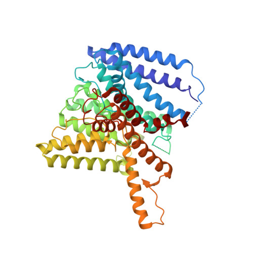
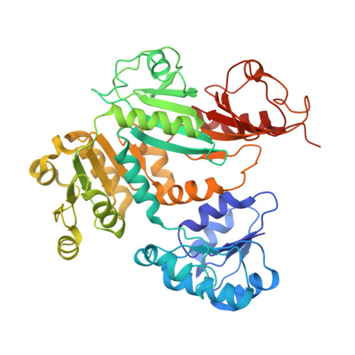
TrkA undergoes a tetramer-to-dimer conversion to open TrkH which enables changes in membrane potential.

Zhang, H.Pan, Y.Hu, L.Hudson, M.A.Hofstetter, K.S.Xu, Z.Rong, M.Wang, Z.Prasad, B.V.V.Lockless, S.W.Chiu, W.Zhou, M.

(2020) Nat Commun 11: 547-547

  • DOI: https://doi.org/10.1038/s41467-019-14240-9
  • Primary Citation of Related Structures:  
    6V4J, 6V4K, 6V4L

  • PubMed Abstract: 

    TrkH is a bacterial ion channel implicated in K + uptake and pH regulation. TrkH assembles with its regulatory protein, TrkA, which closes the channel when bound to ADP and opens it when bound to ATP. However, it is unknown how nucleotides control the gating of TrkH through TrkA. Here we report the structures of the TrkH-TrkA complex in the presence of ADP or ATP. TrkA forms a tetrameric ring when bound to ADP and constrains TrkH to a closed conformation. The TrkA ring splits into two TrkA dimers in the presence of ATP and releases the constraints on TrkH, resulting in an open channel conformation. Functional studies show that both the tetramer-to-dimer conversion of TrkA and the loss of constraints on TrkH are required for channel gating. In addition, deletion of TrkA in Escherichia coli depolarizes the cell, suggesting that the TrkH-TrkA complex couples changes in intracellular nucleotides to membrane potential.


  • Organizational Affiliation

    Verna and Marrs McLean Department of Biochemistry and Molecular Biology, Baylor College of Medicine, Houston, TX, 77030, USA.


Macromolecules
Find similar proteins by:  (by identity cutoff)  |  3D Structure
Entity ID: 1
MoleculeChains Sequence LengthOrganismDetailsImage
Trk system potassium uptake protein
A, B, C, D
485Vibrio parahaemolyticusMutation(s): 0 
Gene Names: ACS91_07345BA740_20060BS585_02545C1S91_23870C9I78_16125CA163_10465CGH73_23020CGJ02_21120FHP20_22005WR32_21470
Membrane Entity: Yes 
UniProt
Find proteins for Q87TN7 (Vibrio parahaemolyticus serotype O3:K6 (strain RIMD 2210633))
Explore Q87TN7 
Go to UniProtKB:  Q87TN7
Entity Groups  
Sequence Clusters30% Identity50% Identity70% Identity90% Identity95% Identity100% Identity
UniProt GroupQ87TN7
Sequence Annotations
Expand
  • Reference Sequence
Find similar proteins by:  (by identity cutoff)  |  3D Structure
Entity ID: 2
MoleculeChains Sequence LengthOrganismDetailsImage
Potassium transporter peripheral membrane component
E, F, G, H
458Vibrio parahaemolyticusMutation(s): 0 
Gene Names: trkAsapGACS91_07035BA740_20405C1S91_23530CGH73_23350CGJ02_20785WR32_21780
UniProt
Find proteins for Q87KD2 (Vibrio parahaemolyticus serotype O3:K6 (strain RIMD 2210633))
Explore Q87KD2 
Go to UniProtKB:  Q87KD2
Entity Groups  
Sequence Clusters30% Identity50% Identity70% Identity90% Identity95% Identity100% Identity
UniProt GroupQ87KD2
Sequence Annotations
Expand
  • Reference Sequence
Experimental Data & Validation

Experimental Data

  • Method: X-RAY DIFFRACTION
  • Resolution: 3.53 Å
  • R-Value Free: 0.291 
  • R-Value Work: 0.250 
  • R-Value Observed: 0.252 
  • Space Group: C 1 2 1
Unit Cell:
Length ( Å )Angle ( ˚ )
a = 262.101α = 90
b = 188.495β = 133.163
c = 187.911γ = 90
Software Package:
Software NamePurpose
PHENIXrefinement
HKL-2000data reduction
HKL-2000data scaling
PHASERphasing

Structure Validation

View Full Validation Report

Currently 6V4K does not have a validation slider image.


Entry History & Funding Information

Deposition Data


Funding OrganizationLocationGrant Number
National Institutes of Health/National Heart, Lung, and Blood Institute (NIH/NHLBI)United StatesHL086392
National Institutes of Health/National Institute of Diabetes and Digestive and Kidney Disease (NIH/NIDDK)United StatesDK122784

Revision History  (Full details and data files)

  • Version 1.0: 2020-02-12
    Type: Initial release
  • Version 1.1: 2023-10-11
    Changes: Data collection, Database references, Refinement description