6V3Z | pdb_00006v3z

Structure of Salmonella enteritidis Sen1395


Experimental Data Snapshot

  • Method: X-RAY DIFFRACTION
  • Resolution: 1.90 Å
  • R-Value Free: 
    0.236 (Depositor), 0.250 (DCC) 
  • R-Value Work: 
    0.201 (Depositor), 0.209 (DCC) 
  • R-Value Observed: 
    0.203 (Depositor) 

Starting Model: experimental
View more details

wwPDB Validation 3D Report Full Report

Validation slider image for 6V3Z

This is version 1.2 of the entry. See complete history

Literature

Mechanisms of substrate recognition by a typhoid toxin secretion-associated muramidase.

Geiger, T.Lara-Tejero, M.Xiong, Y.Galan, J.E.

(2020) Elife 9

  • DOI: https://doi.org/10.7554/eLife.53473
  • Primary Citation Related Structures: 
    6V3Z, 6V40

  • PubMed Abstract: 

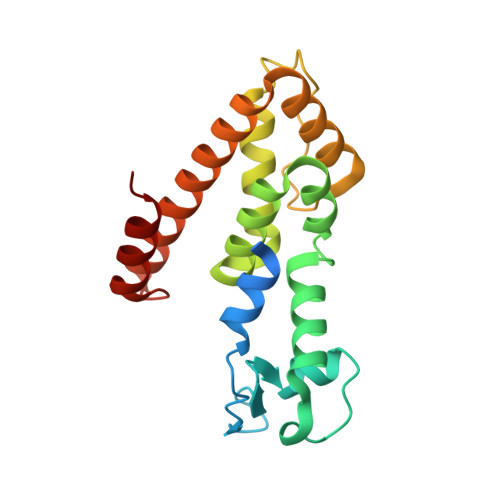
    Typhoid toxin is a virulence factor for the bacterial pathogen Salmonella Typhi, which causes typhoid fever in humans. After its synthesis by intracellular bacteria, typhoid toxin is secreted into the lumen of the Salmonella -containing vacuole by a secretion mechanism strictly dependent on TtsA, a specific muramidase that facilitates toxin transport through the peptidoglycan layer. Here we show that substrate recognition by TtsA depends on a discrete domain within its carboxy terminus, which targets the enzyme to the bacterial poles to recognize YcbB-edited peptidoglycan. Comparison of the atomic structures of TtsA bound to its substrate and that of a close homolog with different specificity identified specific determinants involved in substrate recognition. Combined with structure-guided mutagenesis and in vitro and in vivo crosslinking experiments, this study provides an unprecedented view of the mechanisms by which a muramidase recognizes its peptidoglycan substrate to facilitate protein secretion.


  • Organizational Affiliation
    • Department of Microbial Pathogenesis, Yale University School of Medicine, New Haven, United States.

Macromolecule Content 

  • Total Structure Weight: 44.91 kDa 
  • Atom Count: 2,886 
  • Modeled Residue Count: 356 
  • Deposited Residue Count: 398 
  • Unique protein chains: 1

Macromolecules

Find similar proteins by:|  3D Structure
Entity ID: 1
MoleculeChains  Sequence LengthOrganismDetailsImage
Sen1395
A, B
199Salmonella enterica subsp. enterica serovar EnteritidisMutation(s): 0 
UniProt
Find proteins for A0A1R2HMX1 (Salmonella enteritidis)
Explore A0A1R2HMX1 
Go to UniProtKB:  A0A1R2HMX1
Entity Groups
Sequence Clusters30% Identity50% Identity70% Identity90% Identity95% Identity100% Identity
UniProt GroupA0A1R2HMX1
Sequence Annotations
Expand
Reference Sequence

Experimental Data & Validation

Experimental Data

  • Method: X-RAY DIFFRACTION
  • Resolution: 1.90 Å
  • R-Value Free:  0.236 (Depositor), 0.250 (DCC) 
  • R-Value Work:  0.201 (Depositor), 0.209 (DCC) 
  • R-Value Observed: 0.203 (Depositor) 
Space Group: P 1
Unit Cell:
Length ( Å )Angle ( ˚ )
a = 35.374α = 91.41
b = 49.371β = 107.55
c = 58.639γ = 93.61
Software Package:
Software NamePurpose
REFMACrefinement
PDB_EXTRACTdata extraction
HKL-2000data reduction
HKL-2000data scaling
PHASERphasing

Structure Validation

View Full Validation Report


Entry History 

& Funding Information

Deposition Data


Funding OrganizationLocationGrant Number
National Institutes of Health/National Institute Of Allergy and Infectious Diseases (NIH/NIAID)United States2R01AI114618-06

Revision History  (Full details and data files)

  • Version 1.0: 2020-01-29
    Type: Initial release
  • Version 1.1: 2024-03-06
    Changes: Data collection, Database references, Refinement description
  • Version 1.2: 2024-04-03
    Changes: Refinement description