6V3M | pdb_00006v3m

Crystal structure of 2C-methyl-D-erythritol 2,4-cyclodiphosphate synthase (IspF) Burkholderia pseudomallei in compomplex with ligand HGN-0961 (BSI110840)


Experimental Data Snapshot

  • Method: X-RAY DIFFRACTION
  • Resolution: 1.55 Å
  • R-Value Free: 
    0.169 (Depositor), 0.171 (DCC) 
  • R-Value Work: 
    0.145 (Depositor), 0.146 (DCC) 
  • R-Value Observed: 
    0.145 (Depositor) 

Starting Model: experimental
View more details

wwPDB Validation 3D Report Full Report

Validation slider image for 6V3M

Ligand Structure Quality Assessment 


This is version 1.2 of the entry. See complete history

Literature

Analysis of Burkholderia pseudomallei IspF in complex with sulfapyridine, sulfamonomethoxine, ethoxzolamide and acetazolamide.

Grote, D.Stewart, C.G.Daraji, D.G.Enayati, P.Braverman, K.N.Romanaggi, C.Bolejack, M.J.Yano, J.K.Abendroth, J.Dranow, D.M.Pierce, P.G.Lorimer, D.D.Horanyi, P.S.Staker, B.L.Edwards, T.E.Myler, P.J.Horn, J.R.Hagen, T.J.

(2025) Acta Crystallogr F Struct Biol Commun 81: 138-145

  • DOI: https://doi.org/10.1107/S2053230X25001414
  • Primary Citation Related Structures: 
    6V3M

  • PubMed Abstract: 

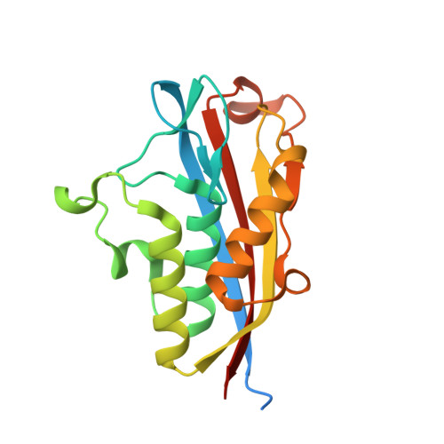
    The methylerythritol phosphate (MEP) pathway is a metabolic pathway that produces the isoprenoids isopentyl pyrophosphate and dimethylallyl pyrophosphate. Notably, the MEP pathway is present in bacteria and not in mammals, which makes the enzymes of the MEP pathway attractive targets for the discovery of new anti-infective agents due to the reduced chances of off-target interactions leading to side effects. There are seven enzymes in the MEP pathway, the fifth of which is IspF. Crystal structures of Burkholderia pseudomallei IspF were determined with five different sulfonamide ligands bound. The sulfonamide-containing ligands were ethoxzolamide, acetazolamide, sulfapyridine and sulfamonomethoxine. The fifth bound ligand was a synthetic analog of acetazolamide. All ligands coordinated to the active-site Zn +2 ion through the sulfonamide group, although sulfapyridine and sulfamonomethoxine, both of which are known antibacterial agents, possess similar binding interactions that are distinct from the other three sulfonamides. These structural data will aid in the discovery of new IspF inhibitors.


  • Organizational Affiliation
    • Department of Chemistry and Biochemistry, Northern Illinois University, 1425 Lincoln Highway, DeKalb, IL 60115, USA.

Macromolecule Content 

  • Total Structure Weight: 60.16 kDa 
  • Atom Count: 4,169 
  • Modeled Residue Count: 480 
  • Deposited Residue Count: 549 
  • Unique protein chains: 1

Macromolecules

Find similar proteins by:|  3D Structure
Entity ID: 1
MoleculeChains  Sequence LengthOrganismDetailsImage
2-C-methyl-D-erythritol 2,4-cyclodiphosphate synthase
A, B, C
183Burkholderia pseudomallei 1710bMutation(s): 0 
Gene Names: ispFBURPS1710b_2511
EC: 4.6.1.12
UniProt
Find proteins for Q3JRA0 (Burkholderia pseudomallei (strain 1710b))
Explore Q3JRA0 
Go to UniProtKB:  Q3JRA0
Entity Groups
Sequence Clusters30% Identity50% Identity70% Identity90% Identity95% Identity100% Identity
UniProt GroupQ3JRA0
Sequence Annotations
Expand
Reference Sequence

Small Molecules

Ligands 5 Unique
IDChains Name / Formula / InChI Key2D Diagram3D Interactions
QOG
(Subject of Investigation/LOI)

Query on QOG


Download:Ideal Coordinates CCD File
E [auth A],
K [auth B],
P [auth C]
5-{[(propan-2-yl)carbamoyl]amino}-1,3,4-thiadiazole-2-sulfonamide
C6 H11 N5 O3 S2
YLZWSQAIKQWBTO-UHFFFAOYSA-N
MLT

Query on MLT


Download:Ideal Coordinates CCD File
I [auth A]D-MALATE
C4 H6 O5
BJEPYKJPYRNKOW-UWTATZPHSA-N
DMS

Query on DMS


Download:Ideal Coordinates CCD File
F [auth A],
G [auth A],
L [auth B],
M [auth B],
Q [auth C]
DIMETHYL SULFOXIDE
C2 H6 O S
IAZDPXIOMUYVGZ-UHFFFAOYSA-N
ZN

Query on ZN


Download:Ideal Coordinates CCD File
D [auth A],
J [auth B],
O [auth C]
ZINC ION
Zn
PTFCDOFLOPIGGS-UHFFFAOYSA-N
EDO

Query on EDO


Download:Ideal Coordinates CCD File
H [auth A],
N [auth B],
R [auth C]
1,2-ETHANEDIOL
C2 H6 O2
LYCAIKOWRPUZTN-UHFFFAOYSA-N

Experimental Data & Validation

Experimental Data

  • Method: X-RAY DIFFRACTION
  • Resolution: 1.55 Å
  • R-Value Free:  0.169 (Depositor), 0.171 (DCC) 
  • R-Value Work:  0.145 (Depositor), 0.146 (DCC) 
  • R-Value Observed: 0.145 (Depositor) 
Space Group: C 1 2 1
Unit Cell:
Length ( Å )Angle ( ˚ )
a = 117.31α = 90
b = 67.55β = 96.317
c = 60.57γ = 90
Software Package:
Software NamePurpose
PHENIXrefinement
XDSdata reduction
XSCALEdata scaling
PDB_EXTRACTdata extraction
PHENIXphasing

Structure Validation

View Full Validation Report


Ligand Structure Quality Assessment 


Entry History 

Deposition Data

Revision History  (Full details and data files)

  • Version 1.0: 2020-12-09
    Type: Initial release
  • Version 1.1: 2023-10-11
    Changes: Data collection, Database references, Refinement description
  • Version 1.2: 2025-04-09
    Changes: Database references, Structure summary