6V37 | pdb_00006v37

K2P2.1(TREK-1)I110D:RuR:ML335 bound channel structure


Experimental Data Snapshot

  • Method: X-RAY DIFFRACTION
  • Resolution: 2.80 Å
  • R-Value Free: 
    0.317 (Depositor), 0.371 (DCC) 
  • R-Value Work: 
    0.272 (Depositor), 0.298 (DCC) 
  • R-Value Observed: 
    0.274 (Depositor) 

Starting Model: experimental
View more details

wwPDB Validation 3D Report Full Report

Validation slider image for 6V37

This is version 1.2 of the entry. See complete history

Literature

Polynuclear Ruthenium Amines Inhibit K2PChannels via a "Finger in the Dam" Mechanism.

Pope, L.Lolicato, M.Minor Jr., D.L.

(2020) Cell Chem Biol 

  • DOI: https://doi.org/10.1016/j.chembiol.2020.01.011
  • Primary Citation Related Structures: 
    6V36, 6V37, 6V3C, 6V3I

  • PubMed Abstract: 

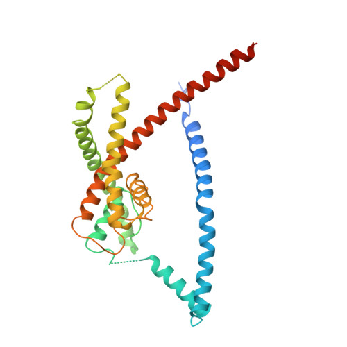
    The trinuclear ruthenium amine ruthenium red (RuR) inhibits diverse ion channels, including K 2P potassium channels, TRPs, the calcium uniporter, CALHMs, ryanodine receptors, and Piezos. Despite this extraordinary array, there is limited information for how RuR engages targets. Here, using X-ray crystallographic and electrophysiological studies of an RuR-sensitive K 2P , K 2P 2.1 (TREK-1) I110D, we show that RuR acts by binding an acidic residue pair comprising the "Keystone inhibitor site" under the K 2P CAP domain archway above the channel pore. We further establish that Ru360, a dinuclear ruthenium amine not known to affect K 2P s, inhibits RuR-sensitive K 2P s using the same mechanism. Structural knowledge enabled a generalizable design strategy for creating K 2P RuR "super-responders" having nanomolar sensitivity. Together, the data define a "finger in the dam" inhibition mechanism acting at a novel K 2P inhibitor binding site. These findings highlight the polysite nature of K 2P pharmacology and provide a new framework for K 2P inhibitor development.


  • Organizational Affiliation
    • Cardiovascular Research Institute, University of California, San Francisco, CA 93858-2330, USA.

Macromolecule Content 

  • Total Structure Weight: 71.22 kDa 
  • Atom Count: 4,309 
  • Modeled Residue Count: 534 
  • Deposited Residue Count: 624 
  • Unique protein chains: 1

Macromolecules

Find similar proteins by:|  3D Structure
Entity ID: 1
MoleculeChains  Sequence LengthOrganismDetailsImage
Potassium channel subfamily K member 2
A, B
312Mus musculusMutation(s): 0 
Membrane Entity: Yes 
UniProt & NIH Common Fund Data Resources
Find proteins for P97438 (Mus musculus)
Explore P97438 
Go to UniProtKB:  P97438
IMPC:  MGI:109366
Entity Groups
Sequence Clusters30% Identity50% Identity70% Identity90% Identity95% Identity100% Identity
UniProt GroupP97438
Sequence Annotations
Expand
Reference Sequence

Small Molecules

Ligands 6 Unique
IDChains Name / Formula / InChI Key2D Diagram3D Interactions
R2R
(Subject of Investigation/LOI)

Query on R2R


Download:Ideal Coordinates CCD File
C [auth A]ruthenium(6+) azanide pentaamino(oxido)ruthenium (1/4/2)
H28 N14 O2 Ru3
FOOPNNMQSUSGAT-UHFFFAOYSA-N
Q6F

Query on Q6F


Download:Ideal Coordinates CCD File
H [auth A],
P [auth B]
N-[(2,4-dichlorophenyl)methyl]-4-[(methylsulfonyl)amino]benzamide
C15 H14 Cl2 N2 O3 S
RDFIQTZRJRVFHK-UHFFFAOYSA-N
D12

Query on D12


Download:Ideal Coordinates CCD File
E [auth A],
F [auth A],
G [auth A],
O [auth B]
DODECANE
C12 H26
SNRUBQQJIBEYMU-UHFFFAOYSA-N
CD

Query on CD


Download:Ideal Coordinates CCD File
D [auth A],
N [auth B],
R [auth B]
CADMIUM ION
Cd
WLZRMCYVCSSEQC-UHFFFAOYSA-N
HEX

Query on HEX


Download:Ideal Coordinates CCD File
M [auth B]HEXANE
C6 H14
VLKZOEOYAKHREP-UHFFFAOYSA-N
K

Query on K


Download:Ideal Coordinates CCD File
I [auth A],
J [auth A],
K [auth A],
L [auth A],
Q [auth B]
POTASSIUM ION
K
NPYPAHLBTDXSSS-UHFFFAOYSA-N

Experimental Data & Validation

Experimental Data

  • Method: X-RAY DIFFRACTION
  • Resolution: 2.80 Å
  • R-Value Free:  0.317 (Depositor), 0.371 (DCC) 
  • R-Value Work:  0.272 (Depositor), 0.298 (DCC) 
  • R-Value Observed: 0.274 (Depositor) 
Space Group: P 21 21 21
Unit Cell:
Length ( Å )Angle ( ˚ )
a = 67.018α = 90
b = 118.735β = 90
c = 129.036γ = 90
Software Package:
Software NamePurpose
PHENIXrefinement
XDSdata reduction
Aimlessdata scaling
PHASERphasing

Structure Validation

View Full Validation Report


Entry History 

& Funding Information

Deposition Data


Funding OrganizationLocationGrant Number
National Institutes of Health/National Library of Medicine (NIH/NLM)United StatesNIH-R01-MH093603

Revision History  (Full details and data files)

  • Version 1.0: 2020-02-26
    Type: Initial release
  • Version 1.1: 2023-10-11
    Changes: Data collection, Database references, Derived calculations, Refinement description
  • Version 1.2: 2024-10-16
    Changes: Structure summary