


Crystal structure of BRAF V600E oncogenic mutant in complex with TAK-580
Gonzalez Del-Pino, G., Li, K., Eck, M.J.To be published.
Experimental Data Snapshot
Starting Model: experimental
View more details
Entity ID: 1 | |||||
|---|---|---|---|---|---|
| Molecule | Chains | Sequence Length | Organism | Details | Image |
| Serine/threonine-protein kinase B-raf | 283 | Homo sapiens | Mutation(s): 15 Gene Names: BRAF EC: 2.7.11.1 | 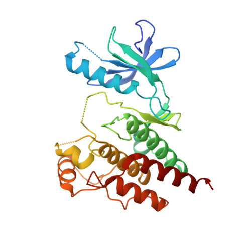 | |
UniProt & NIH Common Fund Data Resources | |||||
PHAROS: P15056 GTEx: ENSG00000157764 | |||||
Entity Groups | |||||
| Sequence Clusters | 30% Identity50% Identity70% Identity90% Identity95% Identity100% Identity | ||||
| UniProt Group | P15056 | ||||
Sequence AnnotationsExpand | |||||
Reference Sequence | |||||
| Ligands 1 Unique | |||||
|---|---|---|---|---|---|
| ID | Chains | Name / Formula / InChI Key | 2D Diagram | 3D Interactions | |
| QOP (Subject of Investigation/LOI) Download:Ideal Coordinates CCD File | C [auth A], D [auth B] | 6-amino-5-chloro-N-[(1R)-1-(5-{[5-chloro-4-(trifluoromethyl)pyridin-2-yl]carbamoyl}-1,3-thiazol-2-yl)ethyl]pyrimidine-4-carboxamide C17 H12 Cl2 F3 N7 O2 S VWMJHAFYPMOMGF-ZCFIWIBFSA-N |  | ||
| Length ( Å ) | Angle ( ˚ ) |
|---|---|
| a = 48.802 | α = 90 |
| b = 85.244 | β = 90 |
| c = 122.905 | γ = 90 |
| Software Name | Purpose |
|---|---|
| PHENIX | refinement |
| PDB_EXTRACT | data extraction |
| XDS | data reduction |
| Aimless | data scaling |
| PHASER | phasing |
| Funding Organization | Location | Grant Number |
|---|---|---|
| National Institutes of Health/National Cancer Institute (NIH/NCI) | United States | P50 CA165962 |
| National Institutes of Health/National Cancer Institute (NIH/NCI) | United States | R35 CA242461 |


