6UZL | pdb_00006uzl

Cryo-EM structure of nucleotide-free MsbA reconstituted into peptidiscs, conformation 2


Experimental Data Snapshot

  • Method: ELECTRON MICROSCOPY
  • Resolution: 4.40 Å
  • Aggregation State: PARTICLE 
  • Reconstruction Method: SINGLE PARTICLE 

wwPDB Validation 3D Report Full Report

Validation slider image for 6UZL

This is version 1.3 of the entry. See complete history

Literature

New approach for membrane protein reconstitution into peptidiscs and basis for their adaptability to different proteins.

Angiulli, G.Dhupar, H.S.Suzuki, H.Wason, I.S.Duong Van Hoa, F.Walz, T.

(2020) Elife 9

  • DOI: https://doi.org/10.7554/eLife.53530
  • Primary Citation Related Structures: 
    6UZ2, 6UZH, 6UZL

  • PubMed Abstract: 

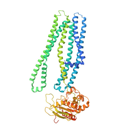
    Previously we introduced peptidiscs as an alternative to detergents to stabilize membrane proteins in solution (Carlson et al., 2018). Here, we present 'on-gradient' reconstitution, a new gentle approach for the reconstitution of labile membrane-protein complexes, and used it to reconstitute Rhodobacter sphaeroides reaction center complexes, demonstrating that peptidiscs can adapt to transmembrane domains of very different sizes and shapes. Using the conventional 'on-bead' approach, we reconstituted Escherichia coli proteins MsbA and MscS and find that peptidiscs stabilize them in their native conformation and allow for high-resolution structure determination by cryo-electron microscopy. The structures reveal that peptidisc peptides can arrange around transmembrane proteins differently, thus revealing the structural basis for why peptidiscs can stabilize such a large variety of membrane proteins. Together, our results establish the gentle and easy-to-use peptidiscs as a potentially universal alternative to detergents as a means to stabilize membrane proteins in solution for structural and functional studies.


  • Organizational Affiliation
    • Laboratory of Molecular Electron Microscopy, Rockefeller University, New York, United States.

Macromolecule Content 

  • Total Structure Weight: 133.67 kDa 
  • Atom Count: 7,008 
  • Modeled Residue Count: 1,062 
  • Deposited Residue Count: 1,206 
  • Unique protein chains: 1

Macromolecules

Find similar proteins by:|  3D Structure
Entity ID: 1
MoleculeChains  Sequence LengthOrganismDetailsImage
Lipid A export ATP-binding/permease protein MsbA
A, B
603Escherichia coliMutation(s): 0 
EC: 7.5.2.6
Membrane Entity: Yes 
UniProt
Find proteins for P60752 (Escherichia coli (strain K12))
Explore P60752 
Go to UniProtKB:  P60752
Entity Groups
Sequence Clusters30% Identity50% Identity70% Identity90% Identity95% Identity100% Identity
UniProt GroupP60752
Sequence Annotations
Expand
Reference Sequence

Experimental Data & Validation

Experimental Data

  • Method: ELECTRON MICROSCOPY
  • Resolution: 4.40 Å
  • Aggregation State: PARTICLE 
  • Reconstruction Method: SINGLE PARTICLE 
EM Software:
TaskSoftware PackageVersion
MODEL REFINEMENTPHENIX
RECONSTRUCTIONRELION3.0

Structure Validation

View Full Validation Report


Entry History 

Deposition Data

Revision History  (Full details and data files)

  • Version 1.0: 2020-03-04
    Type: Initial release
  • Version 1.1: 2020-03-18
    Changes: Database references
  • Version 1.2: 2024-03-06
    Changes: Data collection, Database references
  • Version 1.3: 2025-05-14
    Changes: Data collection, Structure summary