


Structural evidence for a latch mechanism regulating access to the active site of SufS-family cysteine desulfurases
Dunkle, J.A., Bruno, M.R., Frantom, P.A.(2020) Acta Crystallogr D Biol Crystallogr 76: 291-301
Experimental Data Snapshot
Starting Model: experimental
View more details
(2020) Acta Crystallogr D Biol Crystallogr 76: 291-301
Entity ID: 1 | |||||
|---|---|---|---|---|---|
| Molecule | Chains | Sequence Length | Organism | Details | Image |
| Cysteine desulfurase | 406 | Escherichia coli K-12 | Mutation(s): 0 Gene Names: sufS, csdB, ynhB, b1680, JW1670 EC: 2.8.1.7 (PDB Primary Data), 4.4.1.16 (PDB Primary Data), 3.13.1 (UniProt) | 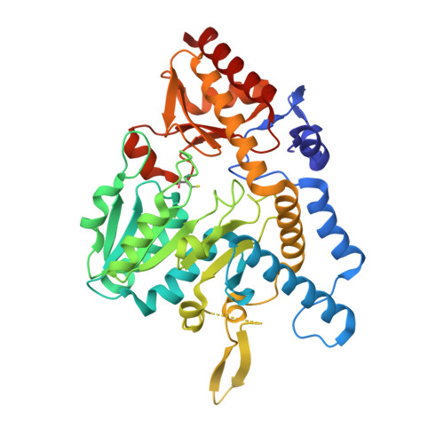 | |
UniProt | |||||
Entity Groups | |||||
| Sequence Clusters | 30% Identity50% Identity70% Identity90% Identity95% Identity100% Identity | ||||
| UniProt Group | P77444 | ||||
Sequence AnnotationsExpand | |||||
Reference Sequence | |||||
| Ligands 1 Unique | |||||
|---|---|---|---|---|---|
| ID | Chains | Name / Formula / InChI Key | 2D Diagram | 3D Interactions | |
| PLP Download:Ideal Coordinates CCD File | C [auth A], D [auth B] | PYRIDOXAL-5'-PHOSPHATE C8 H10 N O6 P NGVDGCNFYWLIFO-UHFFFAOYSA-N |  | ||
| Modified Residues 1 Unique | |||||
|---|---|---|---|---|---|
| ID | Chains | Type | Formula | 2D Diagram | Parent |
| CSS Query on CSS | A, B | L-PEPTIDE LINKING | C3 H7 N O2 S2 |  | CYS |
| Length ( Å ) | Angle ( ˚ ) |
|---|---|
| a = 60.54 | α = 90 |
| b = 115.74 | β = 114.47 |
| c = 63.66 | γ = 90 |
| Software Name | Purpose |
|---|---|
| XDS | data reduction |
| XSCALE | data scaling |
| PHENIX | refinement |
| PDB_EXTRACT | data extraction |
| PHASER | phasing |
| Funding Organization | Location | Grant Number |
|---|---|---|
| National Institutes of Health/National Institute of General Medical Sciences (NIH/NIGMS) | United States | GM112919 |


