6UWF

GltPh in complex with L-aspartate and sodium ions in outward-facing state


Experimental Data Snapshot

  • Method: ELECTRON MICROSCOPY
  • Resolution: 3.08 Å
  • Aggregation State: PARTICLE 
  • Reconstruction Method: SINGLE PARTICLE 

wwPDB Validation   3D Report Full Report


This is version 1.1 of the entry. See complete history


Literature

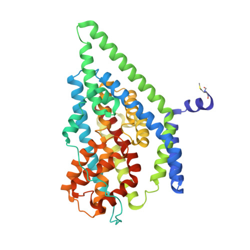
Use of paramagnetic 19 F NMR to monitor domain movement in a glutamate transporter homolog.

Huang, Y.Wang, X.Lv, G.Razavi, A.M.Huysmans, G.H.M.Weinstein, H.Bracken, C.Eliezer, D.Boudker, O.

(2020) Nat Chem Biol 16: 1006-1012

  • DOI: https://doi.org/10.1038/s41589-020-0561-6
  • Primary Citation of Related Structures:  
    6UWF, 6UWL

  • PubMed Abstract: 

    In proteins where conformational changes are functionally important, the number of accessible states and their dynamics are often difficult to establish. Here we describe a novel 19 F-NMR spectroscopy approach to probe dynamics of large membrane proteins. We labeled a glutamate transporter homolog with a 19 F probe via cysteine chemistry and with a Ni 2+ ion via chelation by a di-histidine motif. We used distance-dependent enhancement of the longitudinal relaxation of 19 F nuclei by the paramagnetic metal to assign the observed resonances. We identified one inward- and two outward-facing states of the transporter, in which the substrate-binding site is near the extracellular and intracellular solutions, respectively. We then resolved the structure of the unanticipated second outward-facing state by cryo-EM. Finally, we showed that the rates of the conformational exchange are accessible from measurements of the metal-enhanced longitudinal relaxation of 19 F nuclei.


  • Organizational Affiliation

    Department of Physiology & Biophysics, Weill Cornell Medicine, New York, NY, USA.


Macromolecules
Find similar proteins by:  (by identity cutoff)  |  3D Structure
Entity ID: 1
MoleculeChains Sequence LengthOrganismDetailsImage
Glutamate transporter homolog422Pyrococcus horikoshiiMutation(s): 0 
Membrane Entity: Yes 
UniProt
Find proteins for O59010 (Pyrococcus horikoshii (strain ATCC 700860 / DSM 12428 / JCM 9974 / NBRC 100139 / OT-3))
Explore O59010 
Go to UniProtKB:  O59010
Entity Groups  
Sequence Clusters30% Identity50% Identity70% Identity90% Identity95% Identity100% Identity
UniProt GroupO59010
Sequence Annotations
Expand
  • Reference Sequence
Small Molecules
Ligands 2 Unique
IDChains Name / Formula / InChI Key2D Diagram3D Interactions
6OU
Query on 6OU

Download Ideal Coordinates CCD File 
C [auth A],
D [auth A]
[(2~{R})-1-[2-azanylethoxy(oxidanyl)phosphoryl]oxy-3-hexadecanoyloxy-propan-2-yl] (~{Z})-octadec-9-enoate
C39 H76 N O8 P
FHQVHHIBKUMWTI-OTMQOFQLSA-N
ASP
Query on ASP

Download Ideal Coordinates CCD File 
B [auth A]ASPARTIC ACID
C4 H7 N O4
CKLJMWTZIZZHCS-REOHCLBHSA-N
Modified Residues  1 Unique
IDChains TypeFormula2D DiagramParent
FME
Query on FME
A
L-PEPTIDE LINKINGC6 H11 N O3 SMET
Experimental Data & Validation

Experimental Data

  • Method: ELECTRON MICROSCOPY
  • Resolution: 3.08 Å
  • Aggregation State: PARTICLE 
  • Reconstruction Method: SINGLE PARTICLE 

Structure Validation

View Full Validation Report


Entry History & Funding Information

Deposition Data


Funding OrganizationLocationGrant Number
National Institutes of Health/National Human Genome Research Institute (NIH/NHGRI)United StatesR37NS085318
National Institutes of Health/National Human Genome Research Institute (NIH/NHGRI)United StatesR01NS064357

Revision History  (Full details and data files)

  • Version 1.0: 2020-06-03
    Type: Initial release
  • Version 1.1: 2020-11-18
    Changes: Database references