6URH | pdb_00006urh

Crystal structure of broadly neutralizing antibody AR3X in complex with Hepatitis C virus envelope glycoprotein E2 ectodomain


Experimental Data Snapshot

  • Method: X-RAY DIFFRACTION
  • Resolution: 2.20 Å
  • R-Value Free: 
    0.233 (Depositor), 0.242 (DCC) 
  • R-Value Work: 
    0.186 (Depositor), 0.199 (DCC) 
  • R-Value Observed: 
    0.189 (Depositor) 

Starting Models: experimental
View more details

wwPDB Validation 3D Report Full Report

Validation slider image for 6URH

Ligand Structure Quality Assessment 


This is version 2.2 of the entry. See complete history

Literature

An ultralong CDRH2 in HCV neutralizing antibody demonstrates structural plasticity of antibodies against E2 glycoprotein.

Flyak, A.I.Ruiz, S.E.Salas, J.Rho, S.Bailey, J.R.Bjorkman, P.J.

(2020) Elife 9

  • DOI: https://doi.org/10.7554/eLife.53169
  • Primary Citation Related Structures: 
    6URH

  • PubMed Abstract: 

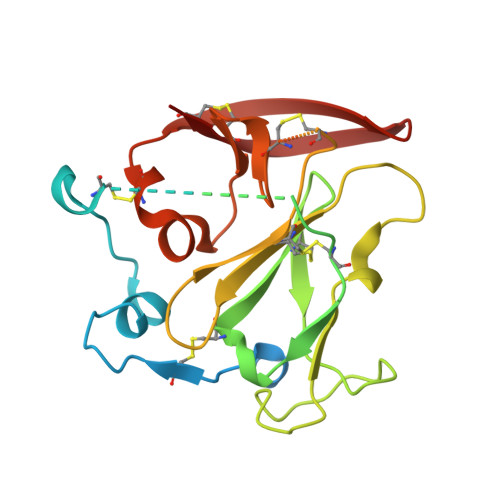
    A vaccine protective against diverse HCV variants is needed to control the HCV epidemic. Structures of E2 complexes with front layer-specific broadly neutralizing antibodies (bNAbs) isolated from HCV-infected individuals, revealed a disulfide bond-containing CDRH3 that adopts straight (individuals who clear infection) or bent (individuals with chronic infection) conformation. To investigate whether a straight versus bent disulfide bond-containing CDRH3 is specific to particular HCV-infected individuals, we solved a crystal structure of the HCV E2 ectodomain in complex with AR3X, a bNAb with an unusually long CDRH2 that was isolated from the chronically-infected individual from whom the bent CDRH3 bNAbs were derived. The structure revealed that AR3X utilizes both its ultralong CDRH2 and a disulfide motif-containing straight CDRH3 to recognize the E2 front layer. These results demonstrate that both the straight and bent CDRH3 classes of HCV bNAb can be elicited in a single individual, revealing a structural plasticity of VH1-69 -derived bNAbs.


  • Organizational Affiliation
    • Division of Biology and Biological Engineering, California Institute of Technology, Pasadena, United States.

Macromolecule Content 

  • Total Structure Weight: 83.12 kDa 
  • Atom Count: 5,124 
  • Modeled Residue Count: 616 
  • Deposited Residue Count: 732 
  • Unique protein chains: 3

Macromolecules

Find similar proteins by:|  3D Structure
Entity ID: 1
MoleculeChains  Sequence LengthOrganismDetailsImage
HCV envelope glycoprotein E2A [auth C]264Orthohepacivirus hominisMutation(s): 0 
UniProt
Find proteins for A0A2P0NE26 (Hepacivirus hominis)
Explore A0A2P0NE26 
Go to UniProtKB:  A0A2P0NE26
Entity Groups
Sequence Clusters30% Identity50% Identity70% Identity90% Identity95% Identity100% Identity
UniProt GroupA0A2P0NE26
Glycosylation
Glycosylation Sites: 5
Sequence Annotations
Expand
Reference Sequence
Find similar proteins by:|  3D Structure
Entity ID: 2
MoleculeChains  Sequence LengthOrganismDetailsImage
AR3X Heavy ChainB [auth H]254Homo sapiensMutation(s): 0 
Find similar proteins by:|  3D Structure
Entity ID: 3
MoleculeChains  Sequence LengthOrganismDetailsImage
AR3X Light ChainC [auth L]214Homo sapiensMutation(s): 0 

Oligosaccharides

Help  
Entity ID: 4
MoleculeChains Length2D Diagram GlycosylationD Interactions
2-acetamido-2-deoxy-beta-D-glucopyranose-(1-4)-2-acetamido-2-deoxy-beta-D-glucopyranoseD [auth A]2N-Glycosylation
Glycosylation Resources
GlyTouCan: G42666HT
GlyCosmos: G42666HT
GlyGen: G42666HT
Entity ID: 5
MoleculeChains Length2D Diagram GlycosylationD Interactions
alpha-D-mannopyranose-(1-6)-beta-D-mannopyranose-(1-3)-2-acetamido-2-deoxy-beta-D-glucopyranose-(1-4)-2-acetamido-2-deoxy-beta-D-glucopyranoseE [auth B]4N-Glycosylation
Glycosylation Resources
GlyTouCan: G12615HA
GlyCosmos: G12615HA
GlyGen: G12615HA
Entity ID: 6
MoleculeChains Length2D Diagram GlycosylationD Interactions
alpha-D-mannopyranose-(1-6)-beta-D-mannopyranose-(1-4)-2-acetamido-2-deoxy-beta-D-glucopyranose-(1-4)-2-acetamido-2-deoxy-beta-D-glucopyranoseF [auth D]4N-Glycosylation
Glycosylation Resources
GlyTouCan: G22573RC
GlyCosmos: G22573RC
GlyGen: G22573RC
Entity ID: 7
MoleculeChains Length2D Diagram GlycosylationD Interactions
alpha-D-mannopyranose-(1-3)-[alpha-D-mannopyranose-(1-6)]alpha-D-mannopyranose-(1-6)-[alpha-D-mannopyranose-(1-3)]beta-D-mannopyranose-(1-4)-2-acetamido-2-deoxy-beta-D-glucopyranose-(1-4)-2-acetamido-2-deoxy-beta-D-glucopyranoseG [auth E]7N-Glycosylation
Glycosylation Resources
GlyTouCan: G55220VL
GlyCosmos: G55220VL
GlyGen: G55220VL

Small Molecules

Ligands 1 Unique
IDChains Name / Formula / InChI Key2D Diagram3D Interactions
NAG

Query on NAG


Download:Ideal Coordinates CCD File
H [auth C]2-acetamido-2-deoxy-beta-D-glucopyranose
C8 H15 N O6
OVRNDRQMDRJTHS-FMDGEEDCSA-N

Experimental Data & Validation

Experimental Data

  • Method: X-RAY DIFFRACTION
  • Resolution: 2.20 Å
  • R-Value Free:  0.233 (Depositor), 0.242 (DCC) 
  • R-Value Work:  0.186 (Depositor), 0.199 (DCC) 
  • R-Value Observed: 0.189 (Depositor) 
Space Group: P 21 21 21
Unit Cell:
Length ( Å )Angle ( ˚ )
a = 64.73α = 90
b = 69.231β = 90
c = 176.699γ = 90
Software Package:
Software NamePurpose
PHENIXrefinement
Aimlessdata scaling
PHASERphasing
PDB_EXTRACTdata extraction
MOSFLMdata reduction

Structure Validation

View Full Validation Report


Ligand Structure Quality Assessment 


Entry History 

& Funding Information

Deposition Data


Funding OrganizationLocationGrant Number
National Institutes of Health/National Institute Of Allergy and Infectious Diseases (NIH/NIAID)United StatesAI127469

Revision History  (Full details and data files)

  • Version 1.0: 2020-03-18
    Type: Initial release
  • Version 2.0: 2020-07-29
    Type: Remediation
    Reason: Carbohydrate remediation
    Changes: Atomic model, Data collection, Derived calculations, Structure summary
  • Version 2.1: 2023-10-11
    Changes: Advisory, Data collection, Database references, Refinement description, Structure summary
  • Version 2.2: 2024-10-30
    Changes: Structure summary