6UOE | pdb_00006uoe

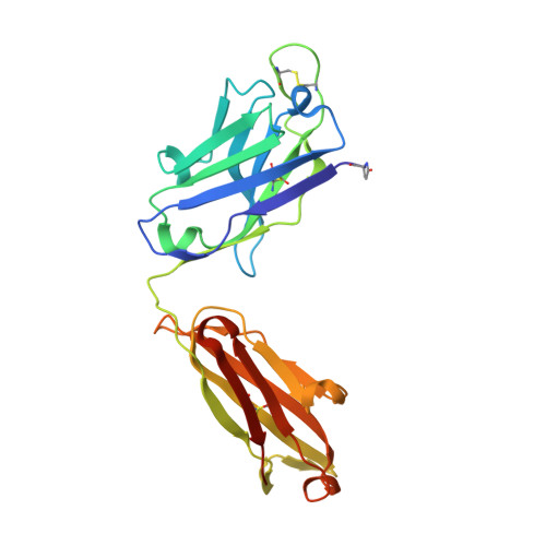
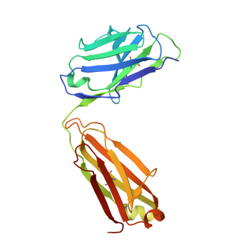
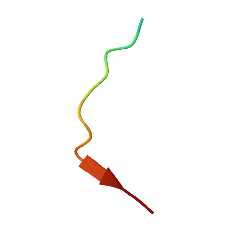
3-25 Fab germline-reversion variant bound to an HCMV gB-derived peptide


Experimental Data Snapshot

  • Method: X-RAY DIFFRACTION
  • Resolution: 1.80 Å
  • R-Value Free: 
    0.186 (Depositor), 0.186 (DCC) 
  • R-Value Work: 
    0.160 (Depositor), 0.162 (DCC) 
  • R-Value Observed: 
    0.162 (Depositor) 

Starting Models: experimental
View more details

wwPDB Validation 3D Report Full Report

Validation slider image for 6UOE

This is version 1.3 of the entry. See complete history. 

Literature

Recognition of a highly conserved glycoprotein B epitope by a bivalent antibody neutralizing HCMV at a post-attachment step.

Ye, X., Su, H., Wrapp, D., Freed, D.C., Li, F., Yuan, Z., Tang, A., Li, L., Ku, Z., Xiong, W., Jaijyan, D., Zhu, H., Wang, D., McLellan, J.S., Zhang, N., Fu, T.M., An, Z.

(2020) PLoS Pathog 16: e1008736-e1008736

  • DOI: https://doi.org/10.1371/journal.ppat.1008736
  • Primary Citation Related Structures: 
    6UOE

  • PubMed Abstract: 

    Human cytomegalovirus (HCMV) is one of the main causative agents of congenital viral infection in neonates. HCMV infection also causes serious morbidity and mortality among organ transplant patients. Glycoprotein B (gB) is a major target for HCMV neutralizing antibodies, yet the underlying neutralization mechanisms remain largely unknown. Here we report that 3-25, a gB-specific monoclonal antibody previously isolated from a healthy HCMV-positive donor, efficiently neutralized 14 HCMV strains in both ARPE-19 cells and MRC-5 cells. The core epitope of 3-25 was mapped to a highly conserved linear epitope on antigenic domain 2 (AD-2) of gB. A 1.8 Å crystal structure of 3-25 Fab in complex with the peptide epitope revealed the molecular determinants of 3-25 binding to gB at atomic resolution. Negative-staining electron microscopy (EM) 3D reconstruction of 3-25 Fab in complex with de-glycosylated postfusion gB showed that 3-25 Fab fully occupied the gB trimer at the N-terminus with flexible binding angles. Functionally, 3-25 efficiently inhibited HCMV infection at a post-attachment step by interfering with viral membrane fusion, and restricted post-infection viral spreading in ARPE-19 cells. Interestingly, bivalency was required for HCMV neutralization by AD-2 specific antibody 3-25 but not the AD-4 specific antibody LJP538. In contrast, bivalency was not required for HCMV binding by both antibodies. Taken together, our results reveal the structural basis of gB recognition by 3-25 and demonstrate that inhibition of viral membrane fusion and a requirement of bivalency may be common for gB AD-2 specific neutralizing antibody.


  • Organizational Affiliation: 
    • Texas Therapeutics Institute, Brown Foundation Institute of Molecular Medicine, University of Texas Health Science Center at Houston, Houston, Texas, United States of America.

Macromolecule Content 

  • Total Structure Weight: 49.91 kDa 
  • Atom Count: 4,091 
  • Modeled Residue Count: 451 
  • Deposited Residue Count: 459 
  • Unique protein chains: 3

Macromolecules

Find similar proteins by:|  3D Structure
Entity ID: 1
MoleculeChains  Sequence LengthOrganismDetailsImage
3-25 Fab heavy chainA [auth H]229Homo sapiensMutation(s): 0 
UniProt
Find proteins for P0DOX5 (Homo sapiens)
Explore P0DOX5 
Go to UniProtKB:  P0DOX5
Entity Groups
Sequence Clusters30% Identity50% Identity70% Identity90% Identity95% Identity100% Identity
UniProt GroupP0DOX5
Sequence Annotations
Expand
Reference Sequence
Find similar proteins by:|  3D Structure
Entity ID: 2
MoleculeChains  Sequence LengthOrganismDetailsImage
3-25 Fab light chainB [auth L]215Homo sapiensMutation(s): 0 
UniProt
Find proteins for P0DOX7 (Homo sapiens)
Explore P0DOX7 
Go to UniProtKB:  P0DOX7
Entity Groups
Sequence Clusters30% Identity50% Identity70% Identity90% Identity95% Identity100% Identity
UniProt GroupP0DOX7
Sequence Annotations
Expand
Reference Sequence
Find similar proteins by:  Sequence   |   3D Structure  
Entity ID: 3
MoleculeChains  Sequence LengthOrganismDetailsImage
Envelope glycoprotein BC [auth P]15Human betaherpesvirus 5Mutation(s): 0 
UniProt
Find proteins for P06473 (Human cytomegalovirus (strain AD169))
Explore P06473 
Go to UniProtKB:  P06473
Entity Groups
Sequence Clusters30% Identity50% Identity70% Identity90% Identity95% Identity100% Identity
UniProt GroupP06473
Sequence Annotations
Expand
Reference Sequence

Small Molecules

Ligands 2 Unique
IDChains Name / Formula / InChI Key2D Diagram3D Interactions
TRS

Query on TRS


Download:Ideal Coordinates CCD File
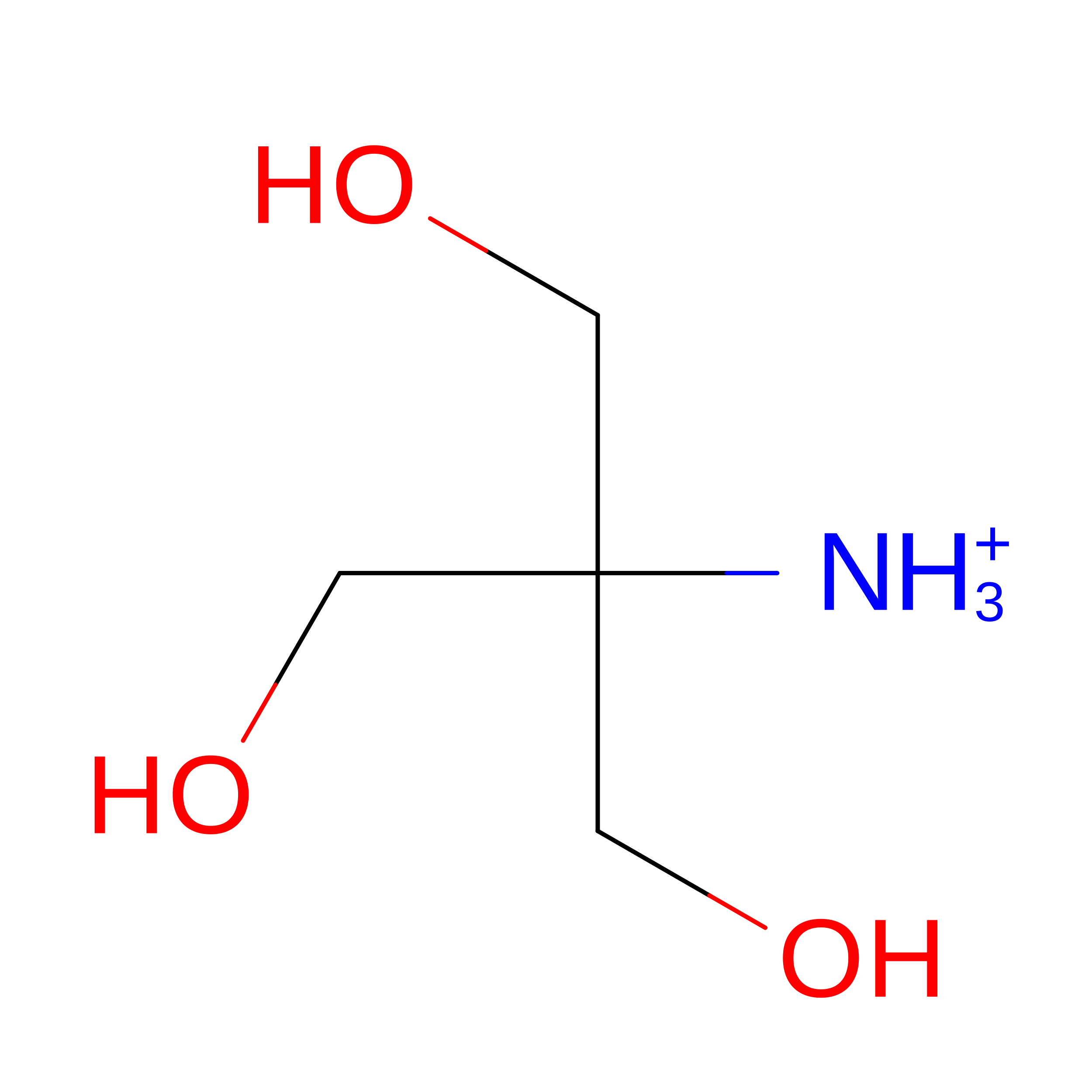
D [auth L]2-AMINO-2-HYDROXYMETHYL-PROPANE-1,3-DIOL
C4 H12 N O3
LENZDBCJOHFCAS-UHFFFAOYSA-O
PEG

Query on PEG


Download:Ideal Coordinates CCD File
E [auth L]DI(HYDROXYETHYL)ETHER
C4 H10 O3
MTHSVFCYNBDYFN-UHFFFAOYSA-N
Modified Residues  1 Unique
IDChains TypeFormula2D DiagramParent
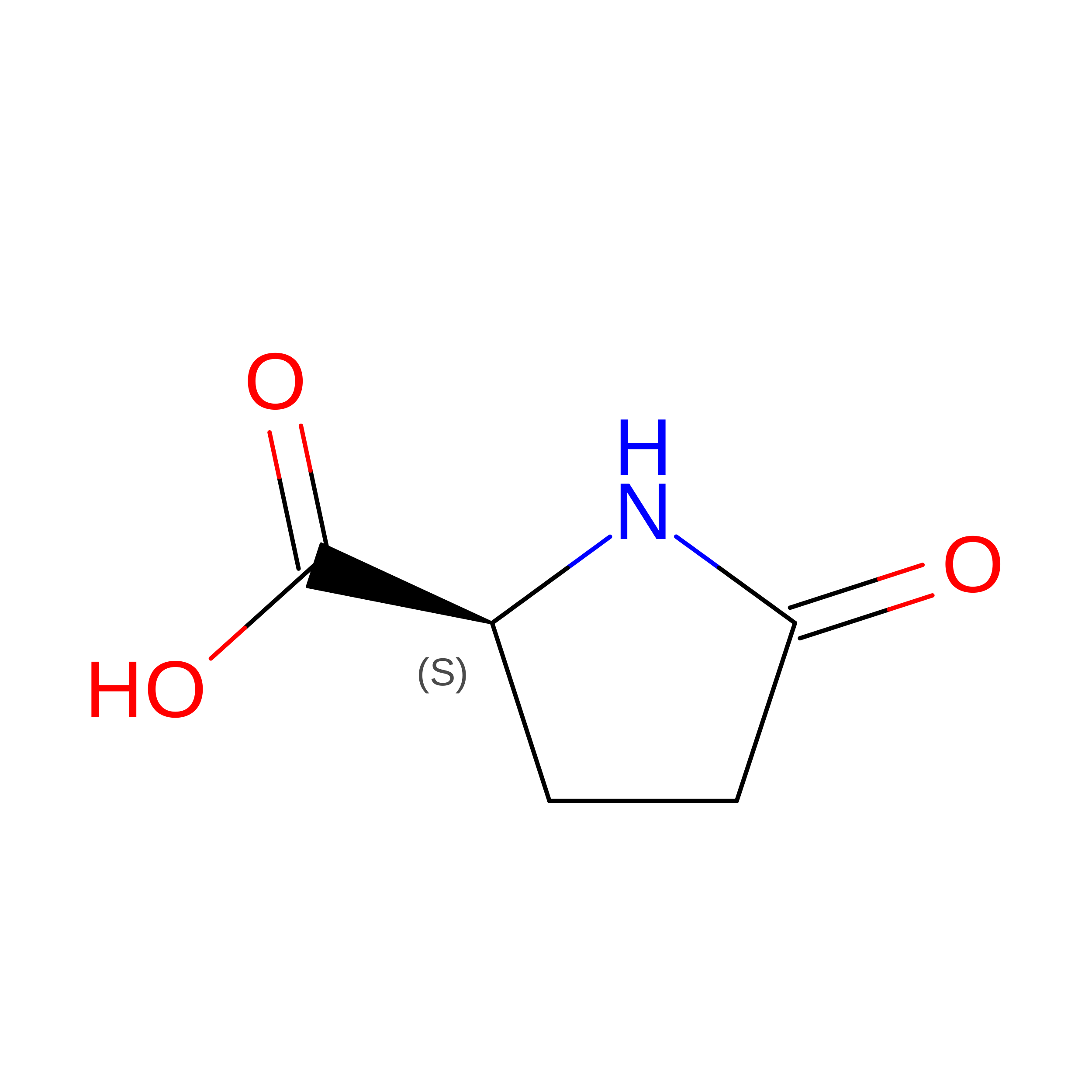
PCA
Query on PCA
A [auth H]L-PEPTIDE LINKINGC5 H7 N O3GLN

Experimental Data & Validation

Experimental Data

  • Method: X-RAY DIFFRACTION
  • Resolution: 1.80 Å
  • R-Value Free:  0.186 (Depositor), 0.186 (DCC) 
  • R-Value Work:  0.160 (Depositor), 0.162 (DCC) 
  • R-Value Observed: 0.162 (Depositor) 
Space Group: C 1 2 1
Unit Cell:
Length ( Å )Angle ( ˚ )
a = 118.663α = 90
b = 67.587β = 133.12
c = 84.031γ = 90
Software Package:
Software NamePurpose
PHENIXrefinement
PDB_EXTRACTdata extraction
MOSFLMdata reduction
Aimlessdata scaling
PHASERphasing

Structure Validation

View Full Validation Report


Entry History 

Deposition Data

Revision History  (Full details and data files)

  • Version 1.0: 2020-07-29
    Type: Initial release
  • Version 1.1: 2021-02-10
    Changes: Database references
  • Version 1.2: 2023-10-11
    Changes: Data collection, Database references, Refinement description
  • Version 1.3: 2024-10-30
    Changes: Structure summary