6UNS

Kinase domain of ALK2-K492A/K493A with LDN-193189


Experimental Data Snapshot

  • Method: X-RAY DIFFRACTION
  • Resolution: 2.30 Å
  • R-Value Free: 0.262 
  • R-Value Work: 0.212 
  • R-Value Observed: 0.215 

wwPDB Validation   3D Report Full Report

Currently 6UNS does not have a validation slider image.


This is version 1.2 of the entry. See complete history


Literature

Structural basis for ALK2/BMPR2 receptor complex signaling through kinase domain oligomerization.

Agnew, C.Ayaz, P.Kashima, R.Loving, H.S.Ghatpande, P.Kung, J.E.Underbakke, E.S.Shan, Y.Shaw, D.E.Hata, A.Jura, N.

(2021) Nat Commun 12: 4950-4950

  • DOI: https://doi.org/10.1038/s41467-021-25248-5
  • Primary Citation of Related Structures:  
    6UNP, 6UNQ, 6UNR, 6UNS

  • PubMed Abstract: 

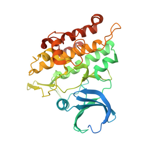
    Upon ligand binding, bone morphogenetic protein (BMP) receptors form active tetrameric complexes, comprised of two type I and two type II receptors, which then transmit signals to SMAD proteins. The link between receptor tetramerization and the mechanism of kinase activation, however, has not been elucidated. Here, using hydrogen deuterium exchange mass spectrometry (HDX-MS), small angle X-ray scattering (SAXS) and molecular dynamics (MD) simulations, combined with analysis of SMAD signaling, we show that the kinase domain of the type I receptor ALK2 and type II receptor BMPR2 form a heterodimeric complex via their C-terminal lobes. Formation of this dimer is essential for ligand-induced receptor signaling and is targeted by mutations in BMPR2 in patients with pulmonary arterial hypertension (PAH). We further show that the type I/type II kinase domain heterodimer serves as the scaffold for assembly of the active tetrameric receptor complexes to enable phosphorylation of the GS domain and activation of SMADs.


  • Organizational Affiliation

    Cardiovascular Research Institute, University of California San Francisco, San Francisco, CA, USA.


Macromolecules
Find similar proteins by:  (by identity cutoff)  |  3D Structure
Entity ID: 1
MoleculeChains Sequence LengthOrganismDetailsImage
Activin receptor type-1
A, B
330Homo sapiensMutation(s): 2 
Gene Names: ACVR1ACVRLK2
EC: 2.7.11.30
UniProt & NIH Common Fund Data Resources
Find proteins for Q04771 (Homo sapiens)
Explore Q04771 
Go to UniProtKB:  Q04771
PHAROS:  Q04771
GTEx:  ENSG00000115170 
Entity Groups  
Sequence Clusters30% Identity50% Identity70% Identity90% Identity95% Identity100% Identity
UniProt GroupQ04771
Sequence Annotations
Expand
  • Reference Sequence
Small Molecules
Ligands 1 Unique
IDChains Name / Formula / InChI Key2D Diagram3D Interactions
LDN
Query on LDN

Download Ideal Coordinates CCD File 
C [auth A],
D [auth B]
4-[6-(4-piperazin-1-ylphenyl)pyrazolo[1,5-a]pyrimidin-3-yl]quinoline
C25 H22 N6
CDOVNWNANFFLFJ-UHFFFAOYSA-N
Experimental Data & Validation

Experimental Data

  • Method: X-RAY DIFFRACTION
  • Resolution: 2.30 Å
  • R-Value Free: 0.262 
  • R-Value Work: 0.212 
  • R-Value Observed: 0.215 
  • Space Group: P 2 21 21
Unit Cell:
Length ( Å )Angle ( ˚ )
a = 60α = 90
b = 83.641β = 90
c = 137.972γ = 90
Software Package:
Software NamePurpose
PHENIXrefinement
XDSdata reduction
Aimlessdata scaling
PHASERphasing

Structure Validation

View Full Validation Report

Currently 6UNS does not have a validation slider image.


Entry History 

Deposition Data

Revision History  (Full details and data files)

  • Version 1.0: 2021-07-07
    Type: Initial release
  • Version 1.1: 2021-10-13
    Changes: Database references
  • Version 1.2: 2023-10-11
    Changes: Data collection, Refinement description